Ronny Schweitzer, Antonio de Marvao, Mit Shah, Paolo Inglese, Peter Kellman, Alaine Berry, Ben Statton, Declan P O'Regan
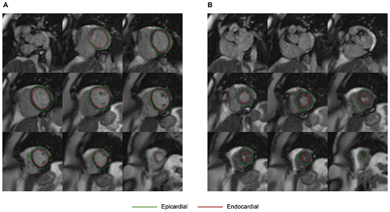
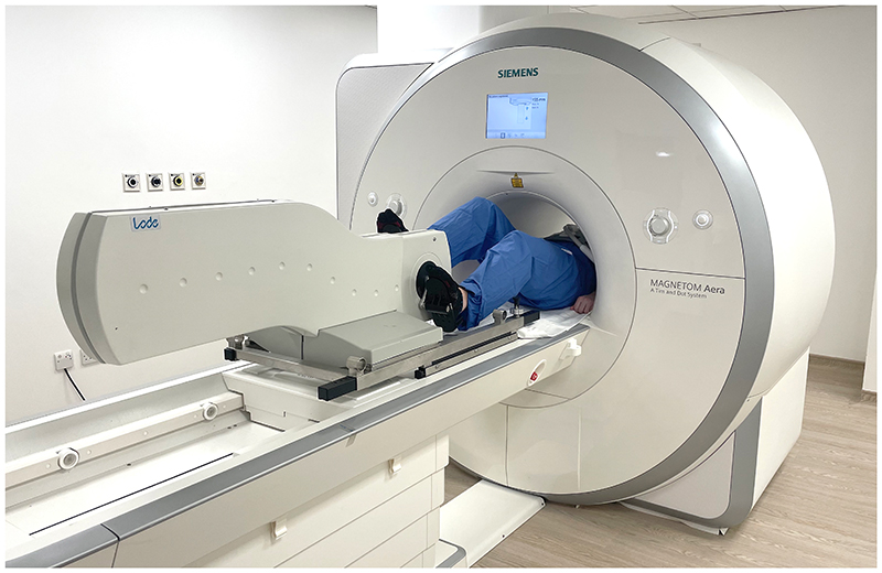
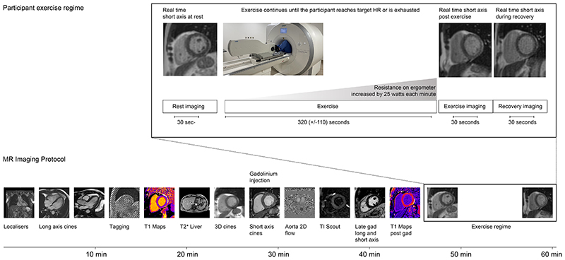
{"title":"建立运动时心血管功能按性别和年龄分层的心脏MRI参考范围。","authors":"Ronny Schweitzer, Antonio de Marvao, Mit Shah, Paolo Inglese, Peter Kellman, Alaine Berry, Ben Statton, Declan P O'Regan","doi":"10.1148/ryct.240175","DOIUrl":null,"url":null,"abstract":"<p><p>Purpose To evaluate the effects of exercise on left ventricular parameters using exercise cardiac MRI in healthy adults without known cardiovascular disease and establish reference ranges stratified by age and sex. Materials and Methods This prospective study included healthy adult participants with no known cardiovascular disease or genetic variants associated with cardiomyopathy, enrolled between January 2018 and April 2021, who underwent exercise cardiac MRI evaluation. Participants were imaged at rest and after exercise, and parameters were measured by two readers. Prediction intervals were calculated and compared across sex and age groups. Results The study included 161 participants (mean age, 49 years ± 14 [SD]; 85 female). Compared with the resting state, exercise caused an increase in heart rate (64 beats per minute ± 9 vs 133 beats per minute ± 19, <i>P</i> < .001), left ventricular end-diastolic volume (140 mL ± 32 vs 148 mL ± 35, <i>P</i> < .001), stroke volume (82 mL ± 18 vs 102 mL ± 25, <i>P</i> < .001), ejection fraction (59% ± 6 vs 69% ± 7, <i>P</i> < .001), and cardiac output (5.2 L/min ± 1.1 vs 13.5 L/min ± 3.9, <i>P</i> < .001) and a decrease in left ventricular end-systolic volume (58 mL ± 18 vs 46 mL ± 15, <i>P</i> < .001). There were statistically significant differences in exercise response between groups stratified by sex and age for most parameters. Conclusion In healthy adults, an increase in cardiac output after exercise was driven by an increase in heart rate with both increased ventricular filling and emptying. Normal ranges for exercise response, stratified by age and sex, were established as a reference for the use of exercise cardiac MRI in clinical practice. <b>Keywords:</b> Cardiac, MR Imaging, Heart, Physiological Studies <i>Supplemental material is available for this article.</i> © RSNA, 2025.</p>","PeriodicalId":21168,"journal":{"name":"Radiology. Cardiothoracic imaging","volume":"7 3","pages":"e240175"},"PeriodicalIF":4.2000,"publicationDate":"2025-06-01","publicationTypes":"Journal Article","fieldsOfStudy":null,"isOpenAccess":false,"openAccessPdf":"https://www.ncbi.nlm.nih.gov/pmc/articles/PMC12207647/pdf/","citationCount":"0","resultStr":"{\"title\":\"Establishing Cardiac MRI Reference Ranges Stratified by Sex and Age for Cardiovascular Function during Exercise.\",\"authors\":\"Ronny Schweitzer, Antonio de Marvao, Mit Shah, Paolo Inglese, Peter Kellman, Alaine Berry, Ben Statton, Declan P O'Regan\",\"doi\":\"10.1148/ryct.240175\",\"DOIUrl\":null,\"url\":null,\"abstract\":\"<p><p>Purpose To evaluate the effects of exercise on left ventricular parameters using exercise cardiac MRI in healthy adults without known cardiovascular disease and establish reference ranges stratified by age and sex. Materials and Methods This prospective study included healthy adult participants with no known cardiovascular disease or genetic variants associated with cardiomyopathy, enrolled between January 2018 and April 2021, who underwent exercise cardiac MRI evaluation. Participants were imaged at rest and after exercise, and parameters were measured by two readers. Prediction intervals were calculated and compared across sex and age groups. Results The study included 161 participants (mean age, 49 years ± 14 [SD]; 85 female). Compared with the resting state, exercise caused an increase in heart rate (64 beats per minute ± 9 vs 133 beats per minute ± 19, <i>P</i> < .001), left ventricular end-diastolic volume (140 mL ± 32 vs 148 mL ± 35, <i>P</i> < .001), stroke volume (82 mL ± 18 vs 102 mL ± 25, <i>P</i> < .001), ejection fraction (59% ± 6 vs 69% ± 7, <i>P</i> < .001), and cardiac output (5.2 L/min ± 1.1 vs 13.5 L/min ± 3.9, <i>P</i> < .001) and a decrease in left ventricular end-systolic volume (58 mL ± 18 vs 46 mL ± 15, <i>P</i> < .001). There were statistically significant differences in exercise response between groups stratified by sex and age for most parameters. Conclusion In healthy adults, an increase in cardiac output after exercise was driven by an increase in heart rate with both increased ventricular filling and emptying. Normal ranges for exercise response, stratified by age and sex, were established as a reference for the use of exercise cardiac MRI in clinical practice. <b>Keywords:</b> Cardiac, MR Imaging, Heart, Physiological Studies <i>Supplemental material is available for this article.</i> © RSNA, 2025.</p>\",\"PeriodicalId\":21168,\"journal\":{\"name\":\"Radiology. Cardiothoracic imaging\",\"volume\":\"7 3\",\"pages\":\"e240175\"},\"PeriodicalIF\":4.2000,\"publicationDate\":\"2025-06-01\",\"publicationTypes\":\"Journal Article\",\"fieldsOfStudy\":null,\"isOpenAccess\":false,\"openAccessPdf\":\"https://www.ncbi.nlm.nih.gov/pmc/articles/PMC12207647/pdf/\",\"citationCount\":\"0\",\"resultStr\":null,\"platform\":\"Semanticscholar\",\"paperid\":null,\"PeriodicalName\":\"Radiology. Cardiothoracic imaging\",\"FirstCategoryId\":\"1085\",\"ListUrlMain\":\"https://doi.org/10.1148/ryct.240175\",\"RegionNum\":0,\"RegionCategory\":null,\"ArticlePicture\":[],\"TitleCN\":null,\"AbstractTextCN\":null,\"PMCID\":null,\"EPubDate\":\"\",\"PubModel\":\"\",\"JCR\":\"Q1\",\"JCRName\":\"RADIOLOGY, NUCLEAR MEDICINE & MEDICAL IMAGING\",\"Score\":null,\"Total\":0}","platform":"Semanticscholar","paperid":null,"PeriodicalName":"Radiology. Cardiothoracic imaging","FirstCategoryId":"1085","ListUrlMain":"https://doi.org/10.1148/ryct.240175","RegionNum":0,"RegionCategory":null,"ArticlePicture":[],"TitleCN":null,"AbstractTextCN":null,"PMCID":null,"EPubDate":"","PubModel":"","JCR":"Q1","JCRName":"RADIOLOGY, NUCLEAR MEDICINE & MEDICAL IMAGING","Score":null,"Total":0}
引用次数: 0

 求助内容:
求助内容: 应助结果提醒方式:


