{"title":"鞍偶发性副神经节瘤1例报告及文献复习。","authors":"Shota Yoshimura, Susumu Yamaguchi, Tomayoshi Hayashi, Takayuki Matsuo","doi":"10.25259/JCIS_140_2024","DOIUrl":null,"url":null,"abstract":"<p><p>Most primary paragangliomas of the head and neck occur in the carotid, jugular body, tympanic ventricle, and vagus nerves. Primary sellar paragangliomas are rare, and their long-term outcomes remain unknown. It is also unclear whether they can be classified as asymptomatic incidentalomas in the sellar region. A 75-year-old man who had been followed up for 15 years for an asymptomatic non-functional pituitary adenoma strongly requested surgery and underwent endoscopic transsphenoidal surgery to remove the tumor. Intraoperatively, the tumor was found to be elastic, harder than the pituitary adenoma, fibrous, and not extremely vascularized. The tumor was excised extracapsularly, although residual tumor tissue remained in the medial part of the bilateral cavernous sinuses. A histopathological assessment revealed negative epithelial markers, positive neuroendocrine markers, and partial positivity for S-100, leading to a diagnosis of paraganglioma. Cervicothoracic and abdominal computed tomography, along with spinal magnetic resonance imaging, revealed no apparent neoplastic lesions. The patient experienced no recurrence for 5 years following the resection. The majority of sellar tumors are pituitary adenomas, craniopharyngiomas, Rathke's cleft cysts, or metastatic brain tumors. Herein, we present a case of an asymptomatic primary sellar paraganglioma that was successfully resected. The case highlights that paraganglioma can be included among incidentalomas in the sellar region. Routine follow-up should generally be recommended for patients with asymptomatic sellar incidentalomas.</p>","PeriodicalId":15512,"journal":{"name":"Journal of Clinical Imaging Science","volume":"15 ","pages":"16"},"PeriodicalIF":1.3000,"publicationDate":"2025-05-07","publicationTypes":"Journal Article","fieldsOfStudy":null,"isOpenAccess":false,"openAccessPdf":"https://www.ncbi.nlm.nih.gov/pmc/articles/PMC12134874/pdf/","citationCount":"0","resultStr":"{\"title\":\"Incidental paraganglioma of sella : A case report and literature review.\",\"authors\":\"Shota Yoshimura, Susumu Yamaguchi, Tomayoshi Hayashi, Takayuki Matsuo\",\"doi\":\"10.25259/JCIS_140_2024\",\"DOIUrl\":null,\"url\":null,\"abstract\":\"<p><p>Most primary paragangliomas of the head and neck occur in the carotid, jugular body, tympanic ventricle, and vagus nerves. Primary sellar paragangliomas are rare, and their long-term outcomes remain unknown. It is also unclear whether they can be classified as asymptomatic incidentalomas in the sellar region. A 75-year-old man who had been followed up for 15 years for an asymptomatic non-functional pituitary adenoma strongly requested surgery and underwent endoscopic transsphenoidal surgery to remove the tumor. Intraoperatively, the tumor was found to be elastic, harder than the pituitary adenoma, fibrous, and not extremely vascularized. The tumor was excised extracapsularly, although residual tumor tissue remained in the medial part of the bilateral cavernous sinuses. A histopathological assessment revealed negative epithelial markers, positive neuroendocrine markers, and partial positivity for S-100, leading to a diagnosis of paraganglioma. Cervicothoracic and abdominal computed tomography, along with spinal magnetic resonance imaging, revealed no apparent neoplastic lesions. The patient experienced no recurrence for 5 years following the resection. The majority of sellar tumors are pituitary adenomas, craniopharyngiomas, Rathke's cleft cysts, or metastatic brain tumors. Herein, we present a case of an asymptomatic primary sellar paraganglioma that was successfully resected. The case highlights that paraganglioma can be included among incidentalomas in the sellar region. Routine follow-up should generally be recommended for patients with asymptomatic sellar incidentalomas.</p>\",\"PeriodicalId\":15512,\"journal\":{\"name\":\"Journal of Clinical Imaging Science\",\"volume\":\"15 \",\"pages\":\"16\"},\"PeriodicalIF\":1.3000,\"publicationDate\":\"2025-05-07\",\"publicationTypes\":\"Journal Article\",\"fieldsOfStudy\":null,\"isOpenAccess\":false,\"openAccessPdf\":\"https://www.ncbi.nlm.nih.gov/pmc/articles/PMC12134874/pdf/\",\"citationCount\":\"0\",\"resultStr\":null,\"platform\":\"Semanticscholar\",\"paperid\":null,\"PeriodicalName\":\"Journal of Clinical Imaging Science\",\"FirstCategoryId\":\"1085\",\"ListUrlMain\":\"https://doi.org/10.25259/JCIS_140_2024\",\"RegionNum\":0,\"RegionCategory\":null,\"ArticlePicture\":[],\"TitleCN\":null,\"AbstractTextCN\":null,\"PMCID\":null,\"EPubDate\":\"2025/1/1 0:00:00\",\"PubModel\":\"eCollection\",\"JCR\":\"Q3\",\"JCRName\":\"RADIOLOGY, NUCLEAR MEDICINE & MEDICAL IMAGING\",\"Score\":null,\"Total\":0}","platform":"Semanticscholar","paperid":null,"PeriodicalName":"Journal of Clinical Imaging Science","FirstCategoryId":"1085","ListUrlMain":"https://doi.org/10.25259/JCIS_140_2024","RegionNum":0,"RegionCategory":null,"ArticlePicture":[],"TitleCN":null,"AbstractTextCN":null,"PMCID":null,"EPubDate":"2025/1/1 0:00:00","PubModel":"eCollection","JCR":"Q3","JCRName":"RADIOLOGY, NUCLEAR MEDICINE & MEDICAL IMAGING","Score":null,"Total":0}
Incidental paraganglioma of sella : A case report and literature review.
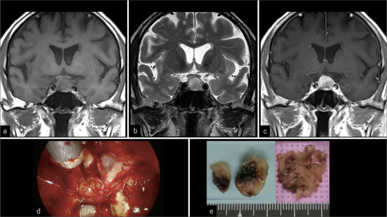
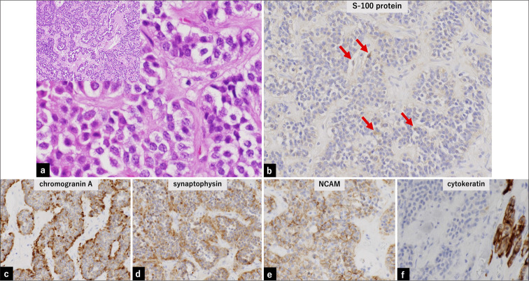
Most primary paragangliomas of the head and neck occur in the carotid, jugular body, tympanic ventricle, and vagus nerves. Primary sellar paragangliomas are rare, and their long-term outcomes remain unknown. It is also unclear whether they can be classified as asymptomatic incidentalomas in the sellar region. A 75-year-old man who had been followed up for 15 years for an asymptomatic non-functional pituitary adenoma strongly requested surgery and underwent endoscopic transsphenoidal surgery to remove the tumor. Intraoperatively, the tumor was found to be elastic, harder than the pituitary adenoma, fibrous, and not extremely vascularized. The tumor was excised extracapsularly, although residual tumor tissue remained in the medial part of the bilateral cavernous sinuses. A histopathological assessment revealed negative epithelial markers, positive neuroendocrine markers, and partial positivity for S-100, leading to a diagnosis of paraganglioma. Cervicothoracic and abdominal computed tomography, along with spinal magnetic resonance imaging, revealed no apparent neoplastic lesions. The patient experienced no recurrence for 5 years following the resection. The majority of sellar tumors are pituitary adenomas, craniopharyngiomas, Rathke's cleft cysts, or metastatic brain tumors. Herein, we present a case of an asymptomatic primary sellar paraganglioma that was successfully resected. The case highlights that paraganglioma can be included among incidentalomas in the sellar region. Routine follow-up should generally be recommended for patients with asymptomatic sellar incidentalomas.
期刊介绍:
The Journal of Clinical Imaging Science (JCIS) is an open access peer-reviewed journal committed to publishing high-quality articles in the field of Imaging Science. The journal aims to present Imaging Science and relevant clinical information in an understandable and useful format. The journal is owned and published by the Scientific Scholar. Audience Our audience includes Radiologists, Researchers, Clinicians, medical professionals and students. Review process JCIS has a highly rigorous peer-review process that makes sure that manuscripts are scientifically accurate, relevant, novel and important. Authors disclose all conflicts, affiliations and financial associations such that the published content is not biased.

 求助内容:
求助内容: 应助结果提醒方式:


