Annette Eidmann, Katharina Kraftborn, Matthias G Walcher, Lukas Fraißler, Maximilian Rudert, Ioannis Stratos
{"title":"踝关节镜融合术后足中部关节的代偿运动影响步态模式。","authors":"Annette Eidmann, Katharina Kraftborn, Matthias G Walcher, Lukas Fraißler, Maximilian Rudert, Ioannis Stratos","doi":"10.1177/24730114251338848","DOIUrl":null,"url":null,"abstract":"<p><strong>Background: </strong>Arthroscopic ankle arthrodesis (AAA) is a standard procedure for end-stage osteoarthritis of the ankle. One of the main concerns after AAA remains the development of secondary osteoarthritis in the subtalar and tarsal joints in the long term. This development is thought to be due to a compensatory increased mobility and therefore increased load on the adjacent joints. Therefore, the aim of the study was to analyze the residual motion of the tarsal joints, the load distribution under the foot, and the influence of tarsal joint motion on load distribution and gait pattern after AAA.</p><p><strong>Methods: </strong>29 patients with arthroscopic AAA were analyzed in a retrospective case-control series by pedobarographic gait analysis and fluoroscopy. The variables examined by pedobarography included peak force, peak pressure, and contact time of 10 different zones of the foot during the roll-over process, comparing the operated with the contralateral healthy foot. The range of motion (ROM) of the subtalar and medial tarsal joints in dorsiflexion/plantarflexion were assessed radiologically.</p><p><strong>Results: </strong>After AAA, peak forces of the ipsilateral foot were significantly reduced for the entire foot and especially the first metatarsal, great toe, and lesser toes during the roll-over process. Peak pressure decreased significantly under the lesser toes and increased under metatarsal 5, without significant load alterations under the mid- and hindfoot. The residual ROM of the subtalar and tarsal joints in dorsiflexion/plantarflexion was 23.5 degrees. The greater the ROM of the adjacent joints, the more the gait pattern normalized.</p><p><strong>Conclusion: </strong>Load distribution during the stance phase is influenced by AAA; the ROM of the subtalar and midfoot joints is essential in normalizing gait pattern.</p><p><strong>Level of evidence: </strong>IV, case series.</p>","PeriodicalId":12429,"journal":{"name":"Foot & Ankle Orthopaedics","volume":"10 2","pages":"24730114251338848"},"PeriodicalIF":0.0000,"publicationDate":"2025-06-03","publicationTypes":"Journal Article","fieldsOfStudy":null,"isOpenAccess":false,"openAccessPdf":"https://www.ncbi.nlm.nih.gov/pmc/articles/PMC12134497/pdf/","citationCount":"0","resultStr":"{\"title\":\"Compensatory Movements of the Midfoot Joints Influence Gait Pattern After Arthroscopic Ankle Arthrodesis.\",\"authors\":\"Annette Eidmann, Katharina Kraftborn, Matthias G Walcher, Lukas Fraißler, Maximilian Rudert, Ioannis Stratos\",\"doi\":\"10.1177/24730114251338848\",\"DOIUrl\":null,\"url\":null,\"abstract\":\"<p><strong>Background: </strong>Arthroscopic ankle arthrodesis (AAA) is a standard procedure for end-stage osteoarthritis of the ankle. One of the main concerns after AAA remains the development of secondary osteoarthritis in the subtalar and tarsal joints in the long term. This development is thought to be due to a compensatory increased mobility and therefore increased load on the adjacent joints. Therefore, the aim of the study was to analyze the residual motion of the tarsal joints, the load distribution under the foot, and the influence of tarsal joint motion on load distribution and gait pattern after AAA.</p><p><strong>Methods: </strong>29 patients with arthroscopic AAA were analyzed in a retrospective case-control series by pedobarographic gait analysis and fluoroscopy. The variables examined by pedobarography included peak force, peak pressure, and contact time of 10 different zones of the foot during the roll-over process, comparing the operated with the contralateral healthy foot. The range of motion (ROM) of the subtalar and medial tarsal joints in dorsiflexion/plantarflexion were assessed radiologically.</p><p><strong>Results: </strong>After AAA, peak forces of the ipsilateral foot were significantly reduced for the entire foot and especially the first metatarsal, great toe, and lesser toes during the roll-over process. Peak pressure decreased significantly under the lesser toes and increased under metatarsal 5, without significant load alterations under the mid- and hindfoot. The residual ROM of the subtalar and tarsal joints in dorsiflexion/plantarflexion was 23.5 degrees. The greater the ROM of the adjacent joints, the more the gait pattern normalized.</p><p><strong>Conclusion: </strong>Load distribution during the stance phase is influenced by AAA; the ROM of the subtalar and midfoot joints is essential in normalizing gait pattern.</p><p><strong>Level of evidence: </strong>IV, case series.</p>\",\"PeriodicalId\":12429,\"journal\":{\"name\":\"Foot & Ankle Orthopaedics\",\"volume\":\"10 2\",\"pages\":\"24730114251338848\"},\"PeriodicalIF\":0.0000,\"publicationDate\":\"2025-06-03\",\"publicationTypes\":\"Journal Article\",\"fieldsOfStudy\":null,\"isOpenAccess\":false,\"openAccessPdf\":\"https://www.ncbi.nlm.nih.gov/pmc/articles/PMC12134497/pdf/\",\"citationCount\":\"0\",\"resultStr\":null,\"platform\":\"Semanticscholar\",\"paperid\":null,\"PeriodicalName\":\"Foot & Ankle Orthopaedics\",\"FirstCategoryId\":\"1085\",\"ListUrlMain\":\"https://doi.org/10.1177/24730114251338848\",\"RegionNum\":0,\"RegionCategory\":null,\"ArticlePicture\":[],\"TitleCN\":null,\"AbstractTextCN\":null,\"PMCID\":null,\"EPubDate\":\"2025/4/1 0:00:00\",\"PubModel\":\"eCollection\",\"JCR\":\"\",\"JCRName\":\"\",\"Score\":null,\"Total\":0}","platform":"Semanticscholar","paperid":null,"PeriodicalName":"Foot & Ankle Orthopaedics","FirstCategoryId":"1085","ListUrlMain":"https://doi.org/10.1177/24730114251338848","RegionNum":0,"RegionCategory":null,"ArticlePicture":[],"TitleCN":null,"AbstractTextCN":null,"PMCID":null,"EPubDate":"2025/4/1 0:00:00","PubModel":"eCollection","JCR":"","JCRName":"","Score":null,"Total":0}
Compensatory Movements of the Midfoot Joints Influence Gait Pattern After Arthroscopic Ankle Arthrodesis.
Background: Arthroscopic ankle arthrodesis (AAA) is a standard procedure for end-stage osteoarthritis of the ankle. One of the main concerns after AAA remains the development of secondary osteoarthritis in the subtalar and tarsal joints in the long term. This development is thought to be due to a compensatory increased mobility and therefore increased load on the adjacent joints. Therefore, the aim of the study was to analyze the residual motion of the tarsal joints, the load distribution under the foot, and the influence of tarsal joint motion on load distribution and gait pattern after AAA.
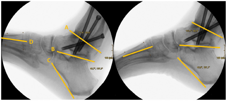
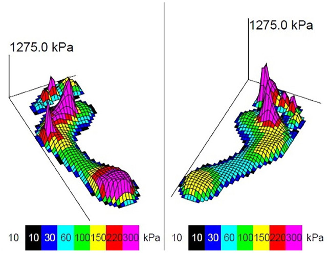
Methods: 29 patients with arthroscopic AAA were analyzed in a retrospective case-control series by pedobarographic gait analysis and fluoroscopy. The variables examined by pedobarography included peak force, peak pressure, and contact time of 10 different zones of the foot during the roll-over process, comparing the operated with the contralateral healthy foot. The range of motion (ROM) of the subtalar and medial tarsal joints in dorsiflexion/plantarflexion were assessed radiologically.
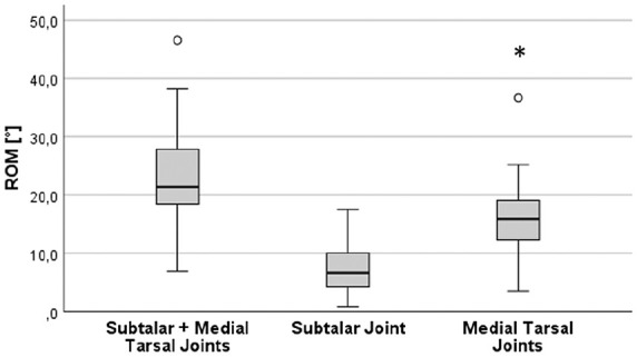
Results: After AAA, peak forces of the ipsilateral foot were significantly reduced for the entire foot and especially the first metatarsal, great toe, and lesser toes during the roll-over process. Peak pressure decreased significantly under the lesser toes and increased under metatarsal 5, without significant load alterations under the mid- and hindfoot. The residual ROM of the subtalar and tarsal joints in dorsiflexion/plantarflexion was 23.5 degrees. The greater the ROM of the adjacent joints, the more the gait pattern normalized.
Conclusion: Load distribution during the stance phase is influenced by AAA; the ROM of the subtalar and midfoot joints is essential in normalizing gait pattern.

 求助内容:
求助内容: 应助结果提醒方式:


