{"title":"不明原因脑膜脑炎犬的脑萎缩","authors":"Rita Gonçalves, Gemma Walmsley, Thomas W. Maddox","doi":"10.1111/jvim.70095","DOIUrl":null,"url":null,"abstract":"<div>\n \n \n <section>\n \n <h3> Background</h3>\n \n <p>Information regarding repeat magnetic resonance imaging (MRI) findings in dogs with meningoencephalitis of unknown origin (MUO) is sparse and it is unknown whether brain atrophy occurs.</p>\n </section>\n \n <section>\n \n <h3> Objectives</h3>\n \n <p>To determine whether brain atrophy occurs in MUO and evaluate if there is an association between atrophy and survival or relapse.</p>\n </section>\n \n <section>\n \n <h3> Animals</h3>\n \n <p>Twenty-three dogs diagnosed with MUO that underwent MRI of the brain on two occasions at least six months apart.</p>\n </section>\n \n <section>\n \n <h3> Methods</h3>\n \n <p>Retrospective study. Interthalamic adhesion thickness to brain height ratio (ITr), lateral ventricle to brain height ratio (LVr), interthalamic adhesion thickness/brain height to lateral ventricle/brain height (ITr/LVr), bicaudate ratio (BCR) and total parenchymal brain volume (TPBV) were measured on both MRI studies and compared.</p>\n </section>\n \n <section>\n \n <h3> Results</h3>\n \n <p>Thirteen dogs relapsed and four died during the study period. Median time between MRIs was 12 months, and only one imaging study (1/23) was considered normal on the second scan. All MRI variables measured significantly changed between imaging studies, but only higher TPBV was associated with increased survival (OR = 1.59, CI = 1.006–2.51, <i>p</i> = 0.047); no variables were found to be associated with relapse. New lesions were identified in six dogs (four of which also showed contrast enhancing lesions), with 5/6 of these dogs subsequently relapsing.</p>\n </section>\n \n <section>\n \n <h3> Conclusions and Clinical Importance</h3>\n \n <p>Brain atrophy likely occurs in dogs with MUO and is associated with worse outcomes. Clinical relapse might be likely in dogs with new or contrast-enhancing lesions on repeat MRI, so more aggressive treatment should be considered in these dogs.</p>\n </section>\n </div>","PeriodicalId":49958,"journal":{"name":"Journal of Veterinary Internal Medicine","volume":"39 4","pages":""},"PeriodicalIF":2.2000,"publicationDate":"2025-06-06","publicationTypes":"Journal Article","fieldsOfStudy":null,"isOpenAccess":false,"openAccessPdf":"https://onlinelibrary.wiley.com/doi/epdf/10.1111/jvim.70095","citationCount":"0","resultStr":"{\"title\":\"Brain Atrophy in Dogs With Meningoencephalitis of Unknown Origin\",\"authors\":\"Rita Gonçalves, Gemma Walmsley, Thomas W. Maddox\",\"doi\":\"10.1111/jvim.70095\",\"DOIUrl\":null,\"url\":null,\"abstract\":\"<div>\\n \\n \\n <section>\\n \\n <h3> Background</h3>\\n \\n <p>Information regarding repeat magnetic resonance imaging (MRI) findings in dogs with meningoencephalitis of unknown origin (MUO) is sparse and it is unknown whether brain atrophy occurs.</p>\\n </section>\\n \\n <section>\\n \\n <h3> Objectives</h3>\\n \\n <p>To determine whether brain atrophy occurs in MUO and evaluate if there is an association between atrophy and survival or relapse.</p>\\n </section>\\n \\n <section>\\n \\n <h3> Animals</h3>\\n \\n <p>Twenty-three dogs diagnosed with MUO that underwent MRI of the brain on two occasions at least six months apart.</p>\\n </section>\\n \\n <section>\\n \\n <h3> Methods</h3>\\n \\n <p>Retrospective study. Interthalamic adhesion thickness to brain height ratio (ITr), lateral ventricle to brain height ratio (LVr), interthalamic adhesion thickness/brain height to lateral ventricle/brain height (ITr/LVr), bicaudate ratio (BCR) and total parenchymal brain volume (TPBV) were measured on both MRI studies and compared.</p>\\n </section>\\n \\n <section>\\n \\n <h3> Results</h3>\\n \\n <p>Thirteen dogs relapsed and four died during the study period. Median time between MRIs was 12 months, and only one imaging study (1/23) was considered normal on the second scan. All MRI variables measured significantly changed between imaging studies, but only higher TPBV was associated with increased survival (OR = 1.59, CI = 1.006–2.51, <i>p</i> = 0.047); no variables were found to be associated with relapse. New lesions were identified in six dogs (four of which also showed contrast enhancing lesions), with 5/6 of these dogs subsequently relapsing.</p>\\n </section>\\n \\n <section>\\n \\n <h3> Conclusions and Clinical Importance</h3>\\n \\n <p>Brain atrophy likely occurs in dogs with MUO and is associated with worse outcomes. Clinical relapse might be likely in dogs with new or contrast-enhancing lesions on repeat MRI, so more aggressive treatment should be considered in these dogs.</p>\\n </section>\\n </div>\",\"PeriodicalId\":49958,\"journal\":{\"name\":\"Journal of Veterinary Internal Medicine\",\"volume\":\"39 4\",\"pages\":\"\"},\"PeriodicalIF\":2.2000,\"publicationDate\":\"2025-06-06\",\"publicationTypes\":\"Journal Article\",\"fieldsOfStudy\":null,\"isOpenAccess\":false,\"openAccessPdf\":\"https://onlinelibrary.wiley.com/doi/epdf/10.1111/jvim.70095\",\"citationCount\":\"0\",\"resultStr\":null,\"platform\":\"Semanticscholar\",\"paperid\":null,\"PeriodicalName\":\"Journal of Veterinary Internal Medicine\",\"FirstCategoryId\":\"97\",\"ListUrlMain\":\"https://onlinelibrary.wiley.com/doi/10.1111/jvim.70095\",\"RegionNum\":2,\"RegionCategory\":\"农林科学\",\"ArticlePicture\":[],\"TitleCN\":null,\"AbstractTextCN\":null,\"PMCID\":null,\"EPubDate\":\"\",\"PubModel\":\"\",\"JCR\":\"Q1\",\"JCRName\":\"VETERINARY SCIENCES\",\"Score\":null,\"Total\":0}","platform":"Semanticscholar","paperid":null,"PeriodicalName":"Journal of Veterinary Internal Medicine","FirstCategoryId":"97","ListUrlMain":"https://onlinelibrary.wiley.com/doi/10.1111/jvim.70095","RegionNum":2,"RegionCategory":"农林科学","ArticlePicture":[],"TitleCN":null,"AbstractTextCN":null,"PMCID":null,"EPubDate":"","PubModel":"","JCR":"Q1","JCRName":"VETERINARY SCIENCES","Score":null,"Total":0}
引用次数: 0
摘要
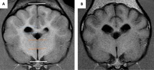
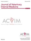
背景关于不明原因脑膜脑炎(MUO)犬的重复磁共振成像(MRI)发现的信息很少,也不清楚是否发生脑萎缩。目的确定MUO患者是否发生脑萎缩,并评估脑萎缩与生存或复发之间是否存在关联。研究人员对23只被诊断患有MUO的狗进行了两次脑部核磁共振检查,时间间隔至少6个月。方法回顾性研究。测量丘脑间黏附厚度与脑高比(ITr)、侧脑室与脑高比(LVr)、丘脑间黏附厚度/脑高与侧脑室/脑高比(ITr/LVr)、双核比(BCR)和脑实质总体积(TPBV),并进行比较。结果研究期间复发犬13只,死亡犬4只。mri之间的中位时间为12个月,只有一次成像研究(1/23)在第二次扫描时被认为是正常的。所有MRI变量在影像学研究之间均有显著变化,但只有较高的TPBV与生存率增加相关(OR = 1.59, CI = 1.006-2.51, p = 0.047);没有发现与复发相关的变量。在6只狗中发现了新的病变(其中4只也显示了对比度增强病变),其中5/6的狗随后复发。结论和临床意义脑萎缩可能发生在患有MUO的狗身上,并与较差的预后相关。在重复MRI上出现新的或增强对比病变的犬可能出现临床复发,因此应考虑对这些犬进行更积极的治疗。
Brain Atrophy in Dogs With Meningoencephalitis of Unknown Origin
Background
Information regarding repeat magnetic resonance imaging (MRI) findings in dogs with meningoencephalitis of unknown origin (MUO) is sparse and it is unknown whether brain atrophy occurs.
Objectives
To determine whether brain atrophy occurs in MUO and evaluate if there is an association between atrophy and survival or relapse.
Animals
Twenty-three dogs diagnosed with MUO that underwent MRI of the brain on two occasions at least six months apart.
Methods
Retrospective study. Interthalamic adhesion thickness to brain height ratio (ITr), lateral ventricle to brain height ratio (LVr), interthalamic adhesion thickness/brain height to lateral ventricle/brain height (ITr/LVr), bicaudate ratio (BCR) and total parenchymal brain volume (TPBV) were measured on both MRI studies and compared.
Results
Thirteen dogs relapsed and four died during the study period. Median time between MRIs was 12 months, and only one imaging study (1/23) was considered normal on the second scan. All MRI variables measured significantly changed between imaging studies, but only higher TPBV was associated with increased survival (OR = 1.59, CI = 1.006–2.51, p = 0.047); no variables were found to be associated with relapse. New lesions were identified in six dogs (four of which also showed contrast enhancing lesions), with 5/6 of these dogs subsequently relapsing.
Conclusions and Clinical Importance
Brain atrophy likely occurs in dogs with MUO and is associated with worse outcomes. Clinical relapse might be likely in dogs with new or contrast-enhancing lesions on repeat MRI, so more aggressive treatment should be considered in these dogs.
期刊介绍:
The mission of the Journal of Veterinary Internal Medicine is to advance veterinary medical knowledge and improve the lives of animals by publication of authoritative scientific articles of animal diseases.
 求助内容:
求助内容: 应助结果提醒方式:


