Abner Velazco, Thomas Glen, Sven Klumpe, Avery Pennington, Jianguo Zhang, Jake L. R. Smith, Calina Glynn, William Bowles, Maryna Kobylynska, Roland A. Fleck, James H. Naismith, Judy S. Kim, Michele C. Darrow, Michael Grange, Angus I. Kirkland, Maud Dumoux
{"title":"原生低温生物样品中SEM电荷伪影的还原","authors":"Abner Velazco, Thomas Glen, Sven Klumpe, Avery Pennington, Jianguo Zhang, Jake L. R. Smith, Calina Glynn, William Bowles, Maryna Kobylynska, Roland A. Fleck, James H. Naismith, Judy S. Kim, Michele C. Darrow, Michael Grange, Angus I. Kirkland, Maud Dumoux","doi":"10.1038/s41467-025-60545-3","DOIUrl":null,"url":null,"abstract":"<p>Scanning electron microscopy (SEM) of frozen-hydrated biological samples allows imaging of subcellular structures at the mesoscale in a representation of their native state. Combined with focused ion beam milling (FIB), serial FIB/SEM can be used to build a 3-dimensional model of cells and tissues. The correlation of specific regions of interest with cryo-electron microscopy (cryoEM) can additionally enable subsequent high-resolution analysis. However, the use of serial FIB/SEM imaging-based methods is often limited due to charging artefacts arising from insulating areas of cryogenically preserved samples. Here, we demonstrate the use of interleaved scanning to attenuate these artefacts, allowing the observation of biological features that otherwise would be masked or distorted. We apply our method to samples where inherent features were not visible using conventional scanning. These examples include membrane contact sites within mammalian cells, visualisation of the degradation compartment in the algae <i>E. gracilis</i> and observation of a network of membranes within different types of axons in an adult mouse cortex. The proposed alternative scanning method could also be applied to imaging other non-conductive specimens in SEM.</p>","PeriodicalId":19066,"journal":{"name":"Nature Communications","volume":"20 1","pages":""},"PeriodicalIF":15.7000,"publicationDate":"2025-06-04","publicationTypes":"Journal Article","fieldsOfStudy":null,"isOpenAccess":false,"openAccessPdf":"","citationCount":"0","resultStr":"{\"title\":\"Reduction of SEM charging artefacts in native cryogenic biological samples\",\"authors\":\"Abner Velazco, Thomas Glen, Sven Klumpe, Avery Pennington, Jianguo Zhang, Jake L. R. Smith, Calina Glynn, William Bowles, Maryna Kobylynska, Roland A. Fleck, James H. Naismith, Judy S. Kim, Michele C. Darrow, Michael Grange, Angus I. Kirkland, Maud Dumoux\",\"doi\":\"10.1038/s41467-025-60545-3\",\"DOIUrl\":null,\"url\":null,\"abstract\":\"<p>Scanning electron microscopy (SEM) of frozen-hydrated biological samples allows imaging of subcellular structures at the mesoscale in a representation of their native state. Combined with focused ion beam milling (FIB), serial FIB/SEM can be used to build a 3-dimensional model of cells and tissues. The correlation of specific regions of interest with cryo-electron microscopy (cryoEM) can additionally enable subsequent high-resolution analysis. However, the use of serial FIB/SEM imaging-based methods is often limited due to charging artefacts arising from insulating areas of cryogenically preserved samples. Here, we demonstrate the use of interleaved scanning to attenuate these artefacts, allowing the observation of biological features that otherwise would be masked or distorted. We apply our method to samples where inherent features were not visible using conventional scanning. These examples include membrane contact sites within mammalian cells, visualisation of the degradation compartment in the algae <i>E. gracilis</i> and observation of a network of membranes within different types of axons in an adult mouse cortex. The proposed alternative scanning method could also be applied to imaging other non-conductive specimens in SEM.</p>\",\"PeriodicalId\":19066,\"journal\":{\"name\":\"Nature Communications\",\"volume\":\"20 1\",\"pages\":\"\"},\"PeriodicalIF\":15.7000,\"publicationDate\":\"2025-06-04\",\"publicationTypes\":\"Journal Article\",\"fieldsOfStudy\":null,\"isOpenAccess\":false,\"openAccessPdf\":\"\",\"citationCount\":\"0\",\"resultStr\":null,\"platform\":\"Semanticscholar\",\"paperid\":null,\"PeriodicalName\":\"Nature Communications\",\"FirstCategoryId\":\"103\",\"ListUrlMain\":\"https://doi.org/10.1038/s41467-025-60545-3\",\"RegionNum\":1,\"RegionCategory\":\"综合性期刊\",\"ArticlePicture\":[],\"TitleCN\":null,\"AbstractTextCN\":null,\"PMCID\":null,\"EPubDate\":\"\",\"PubModel\":\"\",\"JCR\":\"Q1\",\"JCRName\":\"MULTIDISCIPLINARY SCIENCES\",\"Score\":null,\"Total\":0}","platform":"Semanticscholar","paperid":null,"PeriodicalName":"Nature Communications","FirstCategoryId":"103","ListUrlMain":"https://doi.org/10.1038/s41467-025-60545-3","RegionNum":1,"RegionCategory":"综合性期刊","ArticlePicture":[],"TitleCN":null,"AbstractTextCN":null,"PMCID":null,"EPubDate":"","PubModel":"","JCR":"Q1","JCRName":"MULTIDISCIPLINARY SCIENCES","Score":null,"Total":0}
Reduction of SEM charging artefacts in native cryogenic biological samples
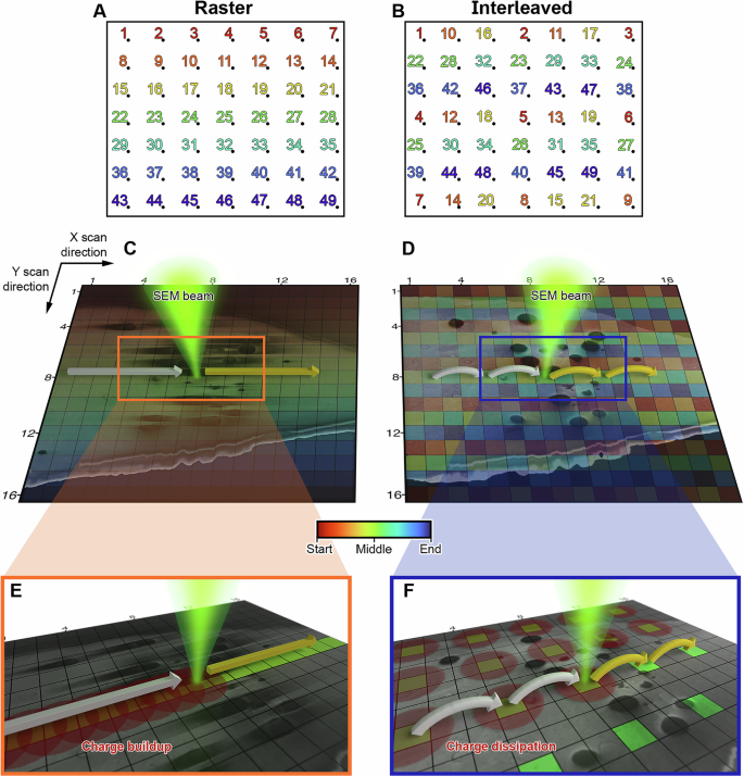
Scanning electron microscopy (SEM) of frozen-hydrated biological samples allows imaging of subcellular structures at the mesoscale in a representation of their native state. Combined with focused ion beam milling (FIB), serial FIB/SEM can be used to build a 3-dimensional model of cells and tissues. The correlation of specific regions of interest with cryo-electron microscopy (cryoEM) can additionally enable subsequent high-resolution analysis. However, the use of serial FIB/SEM imaging-based methods is often limited due to charging artefacts arising from insulating areas of cryogenically preserved samples. Here, we demonstrate the use of interleaved scanning to attenuate these artefacts, allowing the observation of biological features that otherwise would be masked or distorted. We apply our method to samples where inherent features were not visible using conventional scanning. These examples include membrane contact sites within mammalian cells, visualisation of the degradation compartment in the algae E. gracilis and observation of a network of membranes within different types of axons in an adult mouse cortex. The proposed alternative scanning method could also be applied to imaging other non-conductive specimens in SEM.
期刊介绍:
Nature Communications, an open-access journal, publishes high-quality research spanning all areas of the natural sciences. Papers featured in the journal showcase significant advances relevant to specialists in each respective field. With a 2-year impact factor of 16.6 (2022) and a median time of 8 days from submission to the first editorial decision, Nature Communications is committed to rapid dissemination of research findings. As a multidisciplinary journal, it welcomes contributions from biological, health, physical, chemical, Earth, social, mathematical, applied, and engineering sciences, aiming to highlight important breakthroughs within each domain.
 求助内容:
求助内容: 应助结果提醒方式:


