模拟主动脉夹层的主动脉瓣肌腱多模态成像。
IF 1
Q4 CARDIAC & CARDIOVASCULAR SYSTEMS
Journal of Cardiovascular Echography
Pub Date : 2025-01-01
Epub Date: 2025-04-30
DOI:10.4103/jcecho.jcecho_71_24
引用次数: 0
摘要
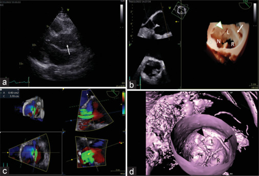
一位53岁的女性被诊断为左心室肥厚。经胸超声心动图显示升主动脉线状密度移动,主动脉偏心不全,提示主动脉夹层。经食管超声心动图和心脏计算机断层扫描(CT)发现复杂的组织带从连接延伸到主动脉壁,证实主动脉瓣肌腱。这是一种极其罕见的主动脉瓣畸形,与主动脉反流有关,类似于主动脉夹层。尽管术前诊断具有挑战性,但三维超声心动图和CT重新格式化有助于空间复杂结构的可视化。本文章由计算机程序翻译,如有差异,请以英文原文为准。
Multimodality Imaging of Aortic Valve Tendon Mimicking Aortic Dissection.
A 53-year-old woman was evaluated for left ventricular hypertrophy. Transthoracic echocardiography showed a mobile linear density in the ascending aorta and eccentric aortic insufficiency, raising the concern of aortic dissection. Transesophageal echocardiography and cardiac computed tomography (CT) found complex bands of tissue extending from the commissure to the aortic wall, confirming aortic valve tendon. It is an extremely rare malformation of aortic valve associated with aortic regurgitation that mimics aortic dissection. Although preoperative diagnosis has been challenging, three-dimensional echocardiography and CT reformatting aided the visualization of the spatially complex structure.
求助全文
通过发布文献求助,成功后即可免费获取论文全文。
去求助
来源期刊

Journal of Cardiovascular Echography
CARDIAC & CARDIOVASCULAR SYSTEMS-
CiteScore
1.40
自引率
12.50%
发文量
27
 求助内容:
求助内容: 应助结果提醒方式:


