Rami Kaddoura, Thuraya Lazkani, Ahmad Abdulhamid Madarati
{"title":"使用3d打印导向和环钻分离式器械进行下颌第一磨牙牙髓显微手术:1例随访2年。","authors":"Rami Kaddoura, Thuraya Lazkani, Ahmad Abdulhamid Madarati","doi":"10.14744/eej.2024.50023","DOIUrl":null,"url":null,"abstract":"<p><p>Endodontic microsurgery (EMS) is a specific treatment modality that targets the root apex of infected teeth that have not been healed by traditional root canal treatments. Recently, the use of advanced three-dimensional (3D) reconstruction technology, such as cone beam computed tomography (CBCT), has improved diagnosis and treatment in dentistry. However, locating the root apex accurately using this technology can be challenging. Also, traditional surgical methods often require significant bone removal which usually results in prolonged surgery and increased risk of trauma and infection. This article introduces the concept of targeted EMS using the 3D-printed surgical guide and a trephine bur to perform single-step osteotomy and root-end resection in complex cases. The surgical guide was designed using a computer-aided planning software to ensure precise angulations and depths of preparation while avoiding critical anatomy regions. The use of the trephine bur enabled efficient and accurate targeted osteotomy regarding the site, angulation, and depth of preparation. This case report describes the use of the 3D-printed guide and the trephine bur to accurately perform EMS of a mandibular first molar with a separated instrument and periapical lesions. (EEJ-2024-03-043).</p>","PeriodicalId":11860,"journal":{"name":"European Endodontic Journal","volume":"10 3","pages":"250-256"},"PeriodicalIF":2.0000,"publicationDate":"2025-05-01","publicationTypes":"Journal Article","fieldsOfStudy":null,"isOpenAccess":false,"openAccessPdf":"https://www.ncbi.nlm.nih.gov/pmc/articles/PMC12102766/pdf/","citationCount":"0","resultStr":"{\"title\":\"Targeted Endodontic Microsurgery of a Mandibular First Molar with a Separated Instrument Using the 3D-printed Guide and Trephine Bur: A Case Report with a 2-year Follow-up.\",\"authors\":\"Rami Kaddoura, Thuraya Lazkani, Ahmad Abdulhamid Madarati\",\"doi\":\"10.14744/eej.2024.50023\",\"DOIUrl\":null,\"url\":null,\"abstract\":\"<p><p>Endodontic microsurgery (EMS) is a specific treatment modality that targets the root apex of infected teeth that have not been healed by traditional root canal treatments. Recently, the use of advanced three-dimensional (3D) reconstruction technology, such as cone beam computed tomography (CBCT), has improved diagnosis and treatment in dentistry. However, locating the root apex accurately using this technology can be challenging. Also, traditional surgical methods often require significant bone removal which usually results in prolonged surgery and increased risk of trauma and infection. This article introduces the concept of targeted EMS using the 3D-printed surgical guide and a trephine bur to perform single-step osteotomy and root-end resection in complex cases. The surgical guide was designed using a computer-aided planning software to ensure precise angulations and depths of preparation while avoiding critical anatomy regions. The use of the trephine bur enabled efficient and accurate targeted osteotomy regarding the site, angulation, and depth of preparation. This case report describes the use of the 3D-printed guide and the trephine bur to accurately perform EMS of a mandibular first molar with a separated instrument and periapical lesions. (EEJ-2024-03-043).</p>\",\"PeriodicalId\":11860,\"journal\":{\"name\":\"European Endodontic Journal\",\"volume\":\"10 3\",\"pages\":\"250-256\"},\"PeriodicalIF\":2.0000,\"publicationDate\":\"2025-05-01\",\"publicationTypes\":\"Journal Article\",\"fieldsOfStudy\":null,\"isOpenAccess\":false,\"openAccessPdf\":\"https://www.ncbi.nlm.nih.gov/pmc/articles/PMC12102766/pdf/\",\"citationCount\":\"0\",\"resultStr\":null,\"platform\":\"Semanticscholar\",\"paperid\":null,\"PeriodicalName\":\"European Endodontic Journal\",\"FirstCategoryId\":\"1085\",\"ListUrlMain\":\"https://doi.org/10.14744/eej.2024.50023\",\"RegionNum\":0,\"RegionCategory\":null,\"ArticlePicture\":[],\"TitleCN\":null,\"AbstractTextCN\":null,\"PMCID\":null,\"EPubDate\":\"\",\"PubModel\":\"\",\"JCR\":\"Q3\",\"JCRName\":\"DENTISTRY, ORAL SURGERY & MEDICINE\",\"Score\":null,\"Total\":0}","platform":"Semanticscholar","paperid":null,"PeriodicalName":"European Endodontic Journal","FirstCategoryId":"1085","ListUrlMain":"https://doi.org/10.14744/eej.2024.50023","RegionNum":0,"RegionCategory":null,"ArticlePicture":[],"TitleCN":null,"AbstractTextCN":null,"PMCID":null,"EPubDate":"","PubModel":"","JCR":"Q3","JCRName":"DENTISTRY, ORAL SURGERY & MEDICINE","Score":null,"Total":0}
Targeted Endodontic Microsurgery of a Mandibular First Molar with a Separated Instrument Using the 3D-printed Guide and Trephine Bur: A Case Report with a 2-year Follow-up.
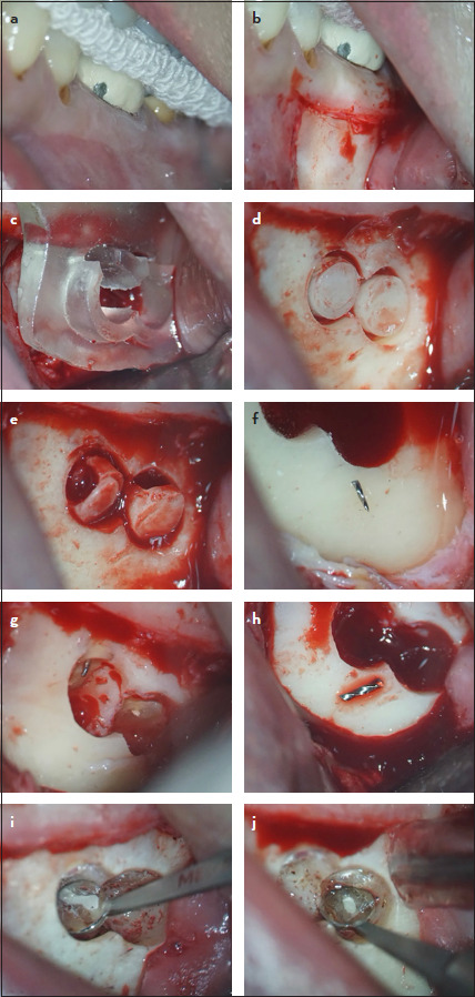
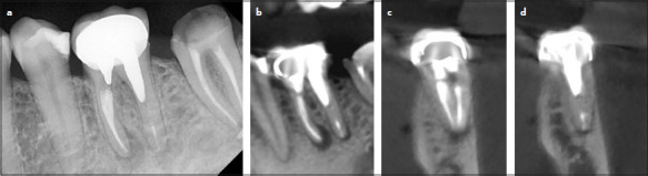
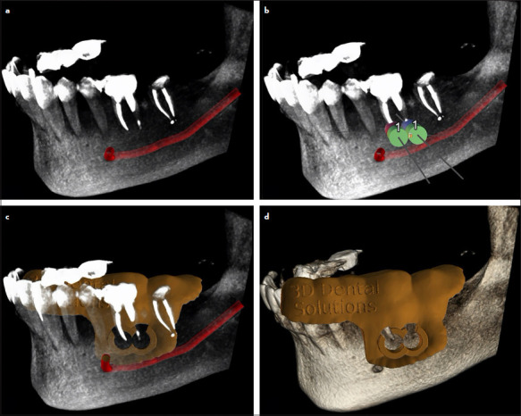
Endodontic microsurgery (EMS) is a specific treatment modality that targets the root apex of infected teeth that have not been healed by traditional root canal treatments. Recently, the use of advanced three-dimensional (3D) reconstruction technology, such as cone beam computed tomography (CBCT), has improved diagnosis and treatment in dentistry. However, locating the root apex accurately using this technology can be challenging. Also, traditional surgical methods often require significant bone removal which usually results in prolonged surgery and increased risk of trauma and infection. This article introduces the concept of targeted EMS using the 3D-printed surgical guide and a trephine bur to perform single-step osteotomy and root-end resection in complex cases. The surgical guide was designed using a computer-aided planning software to ensure precise angulations and depths of preparation while avoiding critical anatomy regions. The use of the trephine bur enabled efficient and accurate targeted osteotomy regarding the site, angulation, and depth of preparation. This case report describes the use of the 3D-printed guide and the trephine bur to accurately perform EMS of a mandibular first molar with a separated instrument and periapical lesions. (EEJ-2024-03-043).

 求助内容:
求助内容: 应助结果提醒方式:


