Hasan Mohsen Al Rammahi, Wen Lin Chai, Hany Mohamed Aly Ahmed
{"title":"使用新型编码系统的马来西亚亚群下颌第一磨牙副管形态:微计算机断层扫描研究。","authors":"Hasan Mohsen Al Rammahi, Wen Lin Chai, Hany Mohamed Aly Ahmed","doi":"10.14744/eej.2025.00922","DOIUrl":null,"url":null,"abstract":"<p><strong>Objective: </strong>This study investigated the morphology of accessory canals in the mandibular first molar of a Malaysian subpopulation.</p><p><strong>Methods: </strong>A total of 140 mandibular first molars were scanned using micro-computed tomography. The accessory canals for each of the mesial and distal roots were classified according to Ahmed et al. system based on location (coronal, middle and apical thirds) and type (patent, blind, loop and delta). A total of thirty mandibular first molar teeth were used for calibration. The Chi-square and Chi-square goodness-fit tests were used to assess the association between the categorical variables. The significance was set at 0.05 (p<0.05).</p><p><strong>Results: </strong>Results showed that the prevalence of accessory canal is 80.71%. The apical third was the most common location for accessory canals in the mesial (79.3%) and distal (75.9%) roots (p<0.001). Amongst accessory canal types, the patent type was the most common (76.43% and 71.43%, respectively) with the codes of M(A1), D(A1). No significant association was found between root type and the presence of the accessory canals (p=0.071). A significant difference was found in the type of accessory canals and the location within the root (p<0.001).</p><p><strong>Conclusion: </strong>In this population, the mandibular first molars showed a high prevalence of accessory canals in the mesial and distal roots. Patent accessory canals in the apical third are the most common in both roots followed by apical delta. (EEJ-2024-10-163).</p>","PeriodicalId":11860,"journal":{"name":"European Endodontic Journal","volume":"10 3","pages":"188-197"},"PeriodicalIF":2.0000,"publicationDate":"2025-05-01","publicationTypes":"Journal Article","fieldsOfStudy":null,"isOpenAccess":false,"openAccessPdf":"https://www.ncbi.nlm.nih.gov/pmc/articles/PMC12102767/pdf/","citationCount":"0","resultStr":"{\"title\":\"Morphology of Accessory Canals in Mandibular First Molar of a Malaysian Subpopulation Using a Novel Coding System: A Micro-computed Tomographic Study.\",\"authors\":\"Hasan Mohsen Al Rammahi, Wen Lin Chai, Hany Mohamed Aly Ahmed\",\"doi\":\"10.14744/eej.2025.00922\",\"DOIUrl\":null,\"url\":null,\"abstract\":\"<p><strong>Objective: </strong>This study investigated the morphology of accessory canals in the mandibular first molar of a Malaysian subpopulation.</p><p><strong>Methods: </strong>A total of 140 mandibular first molars were scanned using micro-computed tomography. The accessory canals for each of the mesial and distal roots were classified according to Ahmed et al. system based on location (coronal, middle and apical thirds) and type (patent, blind, loop and delta). A total of thirty mandibular first molar teeth were used for calibration. The Chi-square and Chi-square goodness-fit tests were used to assess the association between the categorical variables. The significance was set at 0.05 (p<0.05).</p><p><strong>Results: </strong>Results showed that the prevalence of accessory canal is 80.71%. The apical third was the most common location for accessory canals in the mesial (79.3%) and distal (75.9%) roots (p<0.001). Amongst accessory canal types, the patent type was the most common (76.43% and 71.43%, respectively) with the codes of M(A1), D(A1). No significant association was found between root type and the presence of the accessory canals (p=0.071). A significant difference was found in the type of accessory canals and the location within the root (p<0.001).</p><p><strong>Conclusion: </strong>In this population, the mandibular first molars showed a high prevalence of accessory canals in the mesial and distal roots. Patent accessory canals in the apical third are the most common in both roots followed by apical delta. (EEJ-2024-10-163).</p>\",\"PeriodicalId\":11860,\"journal\":{\"name\":\"European Endodontic Journal\",\"volume\":\"10 3\",\"pages\":\"188-197\"},\"PeriodicalIF\":2.0000,\"publicationDate\":\"2025-05-01\",\"publicationTypes\":\"Journal Article\",\"fieldsOfStudy\":null,\"isOpenAccess\":false,\"openAccessPdf\":\"https://www.ncbi.nlm.nih.gov/pmc/articles/PMC12102767/pdf/\",\"citationCount\":\"0\",\"resultStr\":null,\"platform\":\"Semanticscholar\",\"paperid\":null,\"PeriodicalName\":\"European Endodontic Journal\",\"FirstCategoryId\":\"1085\",\"ListUrlMain\":\"https://doi.org/10.14744/eej.2025.00922\",\"RegionNum\":0,\"RegionCategory\":null,\"ArticlePicture\":[],\"TitleCN\":null,\"AbstractTextCN\":null,\"PMCID\":null,\"EPubDate\":\"\",\"PubModel\":\"\",\"JCR\":\"Q3\",\"JCRName\":\"DENTISTRY, ORAL SURGERY & MEDICINE\",\"Score\":null,\"Total\":0}","platform":"Semanticscholar","paperid":null,"PeriodicalName":"European Endodontic Journal","FirstCategoryId":"1085","ListUrlMain":"https://doi.org/10.14744/eej.2025.00922","RegionNum":0,"RegionCategory":null,"ArticlePicture":[],"TitleCN":null,"AbstractTextCN":null,"PMCID":null,"EPubDate":"","PubModel":"","JCR":"Q3","JCRName":"DENTISTRY, ORAL SURGERY & MEDICINE","Score":null,"Total":0}
Morphology of Accessory Canals in Mandibular First Molar of a Malaysian Subpopulation Using a Novel Coding System: A Micro-computed Tomographic Study.
Objective: This study investigated the morphology of accessory canals in the mandibular first molar of a Malaysian subpopulation.
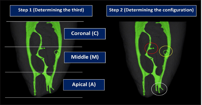
Methods: A total of 140 mandibular first molars were scanned using micro-computed tomography. The accessory canals for each of the mesial and distal roots were classified according to Ahmed et al. system based on location (coronal, middle and apical thirds) and type (patent, blind, loop and delta). A total of thirty mandibular first molar teeth were used for calibration. The Chi-square and Chi-square goodness-fit tests were used to assess the association between the categorical variables. The significance was set at 0.05 (p<0.05).
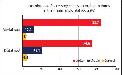
Results: Results showed that the prevalence of accessory canal is 80.71%. The apical third was the most common location for accessory canals in the mesial (79.3%) and distal (75.9%) roots (p<0.001). Amongst accessory canal types, the patent type was the most common (76.43% and 71.43%, respectively) with the codes of M(A1), D(A1). No significant association was found between root type and the presence of the accessory canals (p=0.071). A significant difference was found in the type of accessory canals and the location within the root (p<0.001).
Conclusion: In this population, the mandibular first molars showed a high prevalence of accessory canals in the mesial and distal roots. Patent accessory canals in the apical third are the most common in both roots followed by apical delta. (EEJ-2024-10-163).


 求助内容:
求助内容: 应助结果提醒方式:


