{"title":"异慢性盆腔高级别黏液炎性纤维母细胞肉瘤及子宫内膜样癌伴常见基因突变:罕见病例报告及基因组分析。","authors":"Yuriko Higashi, Mika Mizuno, Ikumi Kitazono, Toshiaki Akahane, Takashi Tasaki, Hirotsugu Noguchi, Masanori Hisaoka, Hiroaki Kobayashi, Akihide Tanimoto","doi":"10.1186/s13000-025-01669-4","DOIUrl":null,"url":null,"abstract":"<p><strong>Objective: </strong>This report presents a rare case involving an extreme epithelial-to-mesenchymal transition, in which a specific type of sarcoma developed heterochronically as a recurrence of endometrioid carcinoma.</p><p><strong>Case presentation: </strong>A female in her 50's presented with abnormal genital bleeding, and an endometrial biopsy revealed endometrioid carcinoma. Following the diagnosis of stage IA endometrioid carcinoma according to the 2008 classification system of the International Federation of Gynecology and Obstetrics, a robot-assisted simple hysterectomy, bilateral salpingo-oophorectomy, and sentinel lymph node navigation surgery were performed. Six months postoperatively, a tumor mass developed in the pelvis. A transrectal needle biopsy revealed spindle cell proliferation, and pelvic tumor resection was conducted for diagnostic therapy. The patient received no adjuvant chemotherapy or radiotherapy after the second surgery and remained free of tumor recurrence for 8 months. The resected yellowish solid tumor mass, measuring 16 × 12 × 9 cm, exhibited hemorrhage, necrosis, and cystic degeneration and was composed of fascicular proliferation of spindle tumor cells showing nuclear pleomorphism and frequent mitotic figures within a myxoid and inflammatory stroma. No epithelial component or organoid patterns were observed. Immunohistochemically, the tumor cells were positive for factor XIIIa, CD10, and cyclin D1, but negative for keratins (AE1/AE3 and CAM5.2) and other specific markers, supporting a diagnosis of high-grade myxoinflammatory fibroblastic sarcoma (MIFS).</p><p><strong>Conclusion: </strong>Genomic analysis revealed identical mutations in PTEN, PIK3R1, CDKN2 A, and TP53 in both the primary uterine endometrioid carcinoma and heterochronic pelvic MIFS. An integrative approach involving histology, immunohistochemistry, and genomic analysis is critical for elucidating the pathogenesis of rare pelvic and uterine tumors.</p>","PeriodicalId":11237,"journal":{"name":"Diagnostic Pathology","volume":"20 1","pages":"71"},"PeriodicalIF":2.3000,"publicationDate":"2025-06-03","publicationTypes":"Journal Article","fieldsOfStudy":null,"isOpenAccess":false,"openAccessPdf":"https://www.ncbi.nlm.nih.gov/pmc/articles/PMC12131376/pdf/","citationCount":"0","resultStr":"{\"title\":\"Heterochronic pelvic high-grade myxoinflammatory fibroblastic sarcoma and uterine endometroid carcinoma harboring common gene mutations: a rare case report with genomic analysis.\",\"authors\":\"Yuriko Higashi, Mika Mizuno, Ikumi Kitazono, Toshiaki Akahane, Takashi Tasaki, Hirotsugu Noguchi, Masanori Hisaoka, Hiroaki Kobayashi, Akihide Tanimoto\",\"doi\":\"10.1186/s13000-025-01669-4\",\"DOIUrl\":null,\"url\":null,\"abstract\":\"<p><strong>Objective: </strong>This report presents a rare case involving an extreme epithelial-to-mesenchymal transition, in which a specific type of sarcoma developed heterochronically as a recurrence of endometrioid carcinoma.</p><p><strong>Case presentation: </strong>A female in her 50's presented with abnormal genital bleeding, and an endometrial biopsy revealed endometrioid carcinoma. Following the diagnosis of stage IA endometrioid carcinoma according to the 2008 classification system of the International Federation of Gynecology and Obstetrics, a robot-assisted simple hysterectomy, bilateral salpingo-oophorectomy, and sentinel lymph node navigation surgery were performed. Six months postoperatively, a tumor mass developed in the pelvis. A transrectal needle biopsy revealed spindle cell proliferation, and pelvic tumor resection was conducted for diagnostic therapy. The patient received no adjuvant chemotherapy or radiotherapy after the second surgery and remained free of tumor recurrence for 8 months. The resected yellowish solid tumor mass, measuring 16 × 12 × 9 cm, exhibited hemorrhage, necrosis, and cystic degeneration and was composed of fascicular proliferation of spindle tumor cells showing nuclear pleomorphism and frequent mitotic figures within a myxoid and inflammatory stroma. No epithelial component or organoid patterns were observed. Immunohistochemically, the tumor cells were positive for factor XIIIa, CD10, and cyclin D1, but negative for keratins (AE1/AE3 and CAM5.2) and other specific markers, supporting a diagnosis of high-grade myxoinflammatory fibroblastic sarcoma (MIFS).</p><p><strong>Conclusion: </strong>Genomic analysis revealed identical mutations in PTEN, PIK3R1, CDKN2 A, and TP53 in both the primary uterine endometrioid carcinoma and heterochronic pelvic MIFS. An integrative approach involving histology, immunohistochemistry, and genomic analysis is critical for elucidating the pathogenesis of rare pelvic and uterine tumors.</p>\",\"PeriodicalId\":11237,\"journal\":{\"name\":\"Diagnostic Pathology\",\"volume\":\"20 1\",\"pages\":\"71\"},\"PeriodicalIF\":2.3000,\"publicationDate\":\"2025-06-03\",\"publicationTypes\":\"Journal Article\",\"fieldsOfStudy\":null,\"isOpenAccess\":false,\"openAccessPdf\":\"https://www.ncbi.nlm.nih.gov/pmc/articles/PMC12131376/pdf/\",\"citationCount\":\"0\",\"resultStr\":null,\"platform\":\"Semanticscholar\",\"paperid\":null,\"PeriodicalName\":\"Diagnostic Pathology\",\"FirstCategoryId\":\"3\",\"ListUrlMain\":\"https://doi.org/10.1186/s13000-025-01669-4\",\"RegionNum\":3,\"RegionCategory\":\"医学\",\"ArticlePicture\":[],\"TitleCN\":null,\"AbstractTextCN\":null,\"PMCID\":null,\"EPubDate\":\"\",\"PubModel\":\"\",\"JCR\":\"Q2\",\"JCRName\":\"PATHOLOGY\",\"Score\":null,\"Total\":0}","platform":"Semanticscholar","paperid":null,"PeriodicalName":"Diagnostic Pathology","FirstCategoryId":"3","ListUrlMain":"https://doi.org/10.1186/s13000-025-01669-4","RegionNum":3,"RegionCategory":"医学","ArticlePicture":[],"TitleCN":null,"AbstractTextCN":null,"PMCID":null,"EPubDate":"","PubModel":"","JCR":"Q2","JCRName":"PATHOLOGY","Score":null,"Total":0}
引用次数: 0
摘要
目的:本报告报告了一例罕见的极端上皮细胞到间质细胞的转移,其中一种特殊类型的肉瘤发展为异长期复发的子宫内膜样癌。病例介绍:一位50多岁的女性,表现为生殖器异常出血,子宫内膜活检显示子宫内膜样癌。根据2008年国际妇产科学联合会(International Federation of Gynecology and Obstetrics)分类系统诊断为IA期子宫内膜样癌后,行机器人辅助单纯子宫切除术、双侧输卵管-卵巢切除术、前哨淋巴结导航手术。术后6个月,骨盆出现肿块。经直肠穿刺活检显示梭形细胞增生,盆腔肿瘤切除进行诊断治疗。患者第二次手术后未接受辅助化疗或放疗,8个月无肿瘤复发。切除的黄色实体瘤块,尺寸为16 × 12 × 9 cm,表现为出血、坏死和囊性变性,由束状增殖的梭形肿瘤细胞组成,在粘液样和炎症间质中表现为核多形性和频繁的有丝分裂象。未观察到上皮成分或类器官。免疫组化结果显示,肿瘤细胞XIIIa因子、CD10和cyclin D1阳性,但角蛋白(AE1/AE3和CAM5.2)和其他特异性标志物阴性,支持高级别黏液炎性纤维母细胞肉瘤(MIFS)的诊断。结论:基因组分析显示PTEN、PIK3R1、cdkn2a和TP53在原发性子宫内膜样癌和异慢性盆腔MIFS中具有相同的突变。包括组织学、免疫组织化学和基因组分析的综合方法对于阐明罕见的盆腔和子宫肿瘤的发病机制至关重要。
Heterochronic pelvic high-grade myxoinflammatory fibroblastic sarcoma and uterine endometroid carcinoma harboring common gene mutations: a rare case report with genomic analysis.
Objective: This report presents a rare case involving an extreme epithelial-to-mesenchymal transition, in which a specific type of sarcoma developed heterochronically as a recurrence of endometrioid carcinoma.
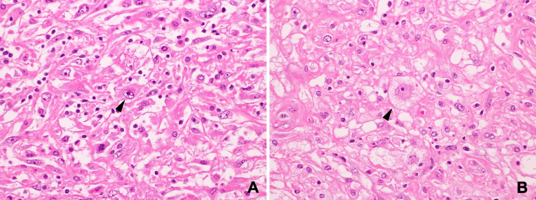
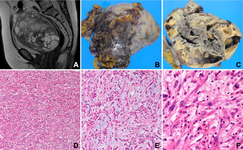
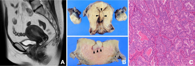
Case presentation: A female in her 50's presented with abnormal genital bleeding, and an endometrial biopsy revealed endometrioid carcinoma. Following the diagnosis of stage IA endometrioid carcinoma according to the 2008 classification system of the International Federation of Gynecology and Obstetrics, a robot-assisted simple hysterectomy, bilateral salpingo-oophorectomy, and sentinel lymph node navigation surgery were performed. Six months postoperatively, a tumor mass developed in the pelvis. A transrectal needle biopsy revealed spindle cell proliferation, and pelvic tumor resection was conducted for diagnostic therapy. The patient received no adjuvant chemotherapy or radiotherapy after the second surgery and remained free of tumor recurrence for 8 months. The resected yellowish solid tumor mass, measuring 16 × 12 × 9 cm, exhibited hemorrhage, necrosis, and cystic degeneration and was composed of fascicular proliferation of spindle tumor cells showing nuclear pleomorphism and frequent mitotic figures within a myxoid and inflammatory stroma. No epithelial component or organoid patterns were observed. Immunohistochemically, the tumor cells were positive for factor XIIIa, CD10, and cyclin D1, but negative for keratins (AE1/AE3 and CAM5.2) and other specific markers, supporting a diagnosis of high-grade myxoinflammatory fibroblastic sarcoma (MIFS).
Conclusion: Genomic analysis revealed identical mutations in PTEN, PIK3R1, CDKN2 A, and TP53 in both the primary uterine endometrioid carcinoma and heterochronic pelvic MIFS. An integrative approach involving histology, immunohistochemistry, and genomic analysis is critical for elucidating the pathogenesis of rare pelvic and uterine tumors.
期刊介绍:
Diagnostic Pathology is an open access, peer-reviewed, online journal that considers research in surgical and clinical pathology, immunology, and biology, with a special focus on cutting-edge approaches in diagnostic pathology and tissue-based therapy. The journal covers all aspects of surgical pathology, including classic diagnostic pathology, prognosis-related diagnosis (tumor stages, prognosis markers, such as MIB-percentage, hormone receptors, etc.), and therapy-related findings. The journal also focuses on the technological aspects of pathology, including molecular biology techniques, morphometry aspects (stereology, DNA analysis, syntactic structure analysis), communication aspects (telecommunication, virtual microscopy, virtual pathology institutions, etc.), and electronic education and quality assurance (for example interactive publication, on-line references with automated updating, etc.).

 求助内容:
求助内容: 应助结果提醒方式:


