Mehdi Dastorani, Mohammad Reza Babaie, Babak Farzaneh, Atousa Haghdoost
{"title":"β - RCS与ADSeal和AH +牙髓密封剂的细胞毒性和皮下组织反应的比较:体外/体内研究","authors":"Mehdi Dastorani, Mohammad Reza Babaie, Babak Farzaneh, Atousa Haghdoost","doi":"10.1007/s44445-025-00013-2","DOIUrl":null,"url":null,"abstract":"<p><p>Sealers are an important part of root canal obturation. Therefore, these materials must be biocompatible and non-toxic to the cells. This study compared the cytotoxicity and tissue response of Beta RCS, ADSeal, and AH Plus. In the in vitro phase, the cytotoxicity of Beta RCS, ADSeal, and AH Plus for human gingival fibroblasts (HGFs) was evaluated using the methyl thiazolyl tetrazolium (MTT) assay after 1, 3, and 7 days (non-toxic defined as > 90% viability). In the in vivo phase, polyethylene tubes containing the sealers (n = 54) and empty control tubes (n = 18) were implanted subcutaneously in 18 Sprague-Dawley rats (4 tubes per rat). The rats were sacrificed after 7, 21 and 42 days. Histological sections were then evaluated under an optical microscope for inflammation, vascular reaction, and fibrous tissue formation. Data were analyzed using the Kruskal-Wallis and Mann-Whitney tests (alpha = 0.05). The cytotoxicity of all three sealers significantly increased with time (P < 0.05). At days 3 and 7, the cytotoxicity of AH Plus was significantly higher than other sealers (P < 0.001). Beta RCS and ADSeal had no significant difference regarding cytotoxicity. At day 21, AH Plus showed significantly higher inflammation and fibrous tissue formation than the control group (P < 0.05). At day 21, tissue reaction around Beta RCS and AH Plus was significantly greater than around ADSeal (P < 0.05). AH Plus showed the highest cytotoxicity while Beta RCS and ADSeal equally had a cytotoxicity comparable to the control group. Both Beta RCS and AH Plus showed high tissue response. ADSeal showed the lowest cytotoxicity and tissue response.</p>","PeriodicalId":47246,"journal":{"name":"Saudi Dental Journal","volume":"37 4-6","pages":"13"},"PeriodicalIF":2.3000,"publicationDate":"2025-06-03","publicationTypes":"Journal Article","fieldsOfStudy":null,"isOpenAccess":false,"openAccessPdf":"https://www.ncbi.nlm.nih.gov/pmc/articles/PMC12129880/pdf/","citationCount":"0","resultStr":"{\"title\":\"Cytotoxicity and subcutaneous tissue response of beta RCS in comparison with ADSeal and AH plus endodontic sealers: in vitro/in vivo study.\",\"authors\":\"Mehdi Dastorani, Mohammad Reza Babaie, Babak Farzaneh, Atousa Haghdoost\",\"doi\":\"10.1007/s44445-025-00013-2\",\"DOIUrl\":null,\"url\":null,\"abstract\":\"<p><p>Sealers are an important part of root canal obturation. Therefore, these materials must be biocompatible and non-toxic to the cells. This study compared the cytotoxicity and tissue response of Beta RCS, ADSeal, and AH Plus. In the in vitro phase, the cytotoxicity of Beta RCS, ADSeal, and AH Plus for human gingival fibroblasts (HGFs) was evaluated using the methyl thiazolyl tetrazolium (MTT) assay after 1, 3, and 7 days (non-toxic defined as > 90% viability). In the in vivo phase, polyethylene tubes containing the sealers (n = 54) and empty control tubes (n = 18) were implanted subcutaneously in 18 Sprague-Dawley rats (4 tubes per rat). The rats were sacrificed after 7, 21 and 42 days. Histological sections were then evaluated under an optical microscope for inflammation, vascular reaction, and fibrous tissue formation. Data were analyzed using the Kruskal-Wallis and Mann-Whitney tests (alpha = 0.05). The cytotoxicity of all three sealers significantly increased with time (P < 0.05). At days 3 and 7, the cytotoxicity of AH Plus was significantly higher than other sealers (P < 0.001). Beta RCS and ADSeal had no significant difference regarding cytotoxicity. At day 21, AH Plus showed significantly higher inflammation and fibrous tissue formation than the control group (P < 0.05). At day 21, tissue reaction around Beta RCS and AH Plus was significantly greater than around ADSeal (P < 0.05). AH Plus showed the highest cytotoxicity while Beta RCS and ADSeal equally had a cytotoxicity comparable to the control group. Both Beta RCS and AH Plus showed high tissue response. ADSeal showed the lowest cytotoxicity and tissue response.</p>\",\"PeriodicalId\":47246,\"journal\":{\"name\":\"Saudi Dental Journal\",\"volume\":\"37 4-6\",\"pages\":\"13\"},\"PeriodicalIF\":2.3000,\"publicationDate\":\"2025-06-03\",\"publicationTypes\":\"Journal Article\",\"fieldsOfStudy\":null,\"isOpenAccess\":false,\"openAccessPdf\":\"https://www.ncbi.nlm.nih.gov/pmc/articles/PMC12129880/pdf/\",\"citationCount\":\"0\",\"resultStr\":null,\"platform\":\"Semanticscholar\",\"paperid\":null,\"PeriodicalName\":\"Saudi Dental Journal\",\"FirstCategoryId\":\"1085\",\"ListUrlMain\":\"https://doi.org/10.1007/s44445-025-00013-2\",\"RegionNum\":0,\"RegionCategory\":null,\"ArticlePicture\":[],\"TitleCN\":null,\"AbstractTextCN\":null,\"PMCID\":null,\"EPubDate\":\"\",\"PubModel\":\"\",\"JCR\":\"Q3\",\"JCRName\":\"DENTISTRY, ORAL SURGERY & MEDICINE\",\"Score\":null,\"Total\":0}","platform":"Semanticscholar","paperid":null,"PeriodicalName":"Saudi Dental Journal","FirstCategoryId":"1085","ListUrlMain":"https://doi.org/10.1007/s44445-025-00013-2","RegionNum":0,"RegionCategory":null,"ArticlePicture":[],"TitleCN":null,"AbstractTextCN":null,"PMCID":null,"EPubDate":"","PubModel":"","JCR":"Q3","JCRName":"DENTISTRY, ORAL SURGERY & MEDICINE","Score":null,"Total":0}
Cytotoxicity and subcutaneous tissue response of beta RCS in comparison with ADSeal and AH plus endodontic sealers: in vitro/in vivo study.
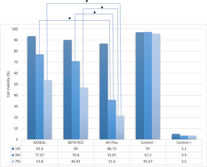
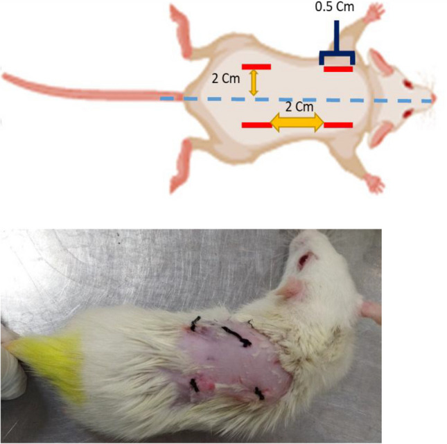
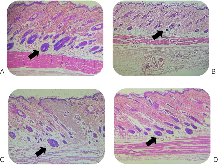
Sealers are an important part of root canal obturation. Therefore, these materials must be biocompatible and non-toxic to the cells. This study compared the cytotoxicity and tissue response of Beta RCS, ADSeal, and AH Plus. In the in vitro phase, the cytotoxicity of Beta RCS, ADSeal, and AH Plus for human gingival fibroblasts (HGFs) was evaluated using the methyl thiazolyl tetrazolium (MTT) assay after 1, 3, and 7 days (non-toxic defined as > 90% viability). In the in vivo phase, polyethylene tubes containing the sealers (n = 54) and empty control tubes (n = 18) were implanted subcutaneously in 18 Sprague-Dawley rats (4 tubes per rat). The rats were sacrificed after 7, 21 and 42 days. Histological sections were then evaluated under an optical microscope for inflammation, vascular reaction, and fibrous tissue formation. Data were analyzed using the Kruskal-Wallis and Mann-Whitney tests (alpha = 0.05). The cytotoxicity of all three sealers significantly increased with time (P < 0.05). At days 3 and 7, the cytotoxicity of AH Plus was significantly higher than other sealers (P < 0.001). Beta RCS and ADSeal had no significant difference regarding cytotoxicity. At day 21, AH Plus showed significantly higher inflammation and fibrous tissue formation than the control group (P < 0.05). At day 21, tissue reaction around Beta RCS and AH Plus was significantly greater than around ADSeal (P < 0.05). AH Plus showed the highest cytotoxicity while Beta RCS and ADSeal equally had a cytotoxicity comparable to the control group. Both Beta RCS and AH Plus showed high tissue response. ADSeal showed the lowest cytotoxicity and tissue response.
期刊介绍:
Saudi Dental Journal is an English language, peer-reviewed scholarly publication in the area of dentistry. Saudi Dental Journal publishes original research and reviews on, but not limited to: • dental disease • clinical trials • dental equipment • new and experimental techniques • epidemiology and oral health • restorative dentistry • periodontology • endodontology • prosthodontics • paediatric dentistry • orthodontics and dental education Saudi Dental Journal is the official publication of the Saudi Dental Society and is published by King Saud University in collaboration with Elsevier and is edited by an international group of eminent researchers.
 求助内容:
求助内容: 应助结果提醒方式:


