{"title":"多中心Castleman病伴淋巴结和皮肤累及的18F-FDG PET/CT","authors":"Merve Yücel, Tarık Şengöz, Nilay Şen Türk","doi":"10.4274/mirt.galenos.2024.80388","DOIUrl":null,"url":null,"abstract":"<p><p>Herein, we describe a rare case of multicentric Castleman disease with multiple lymph node and skin involvement. Ultrasonography of a 38-year-old patient with weakness and fever revealed multiple lymphadenopathies in both inguinal regions. Diagnosed via lymph node biopsy was Castleman's disease, a plasma cell variant. He was diagnosed with prurigo nodularis, lymphocytic vasculitis, and stasis dermatitis in the biopsies of skin lesions located in different regions. <sup>18</sup>F-fluorodeoxyglucose positron emission tomography/computed tomography showed multiple hypermetabolic lymph nodes in the axilla, abdomen, pelvis, and both popliteal areas, multiple hypermetabolic skin thickenings, and skin lesions in both arms, legs, and feet.</p>","PeriodicalId":44681,"journal":{"name":"Molecular Imaging and Radionuclide Therapy","volume":"34 2","pages":"135-138"},"PeriodicalIF":1.1000,"publicationDate":"2025-06-03","publicationTypes":"Journal Article","fieldsOfStudy":null,"isOpenAccess":false,"openAccessPdf":"https://www.ncbi.nlm.nih.gov/pmc/articles/PMC12134962/pdf/","citationCount":"0","resultStr":"{\"title\":\"<sup>18</sup>F-FDG PET/CT of a Multicentric Castleman Disease with Lymph Node and Skin Involvement.\",\"authors\":\"Merve Yücel, Tarık Şengöz, Nilay Şen Türk\",\"doi\":\"10.4274/mirt.galenos.2024.80388\",\"DOIUrl\":null,\"url\":null,\"abstract\":\"<p><p>Herein, we describe a rare case of multicentric Castleman disease with multiple lymph node and skin involvement. Ultrasonography of a 38-year-old patient with weakness and fever revealed multiple lymphadenopathies in both inguinal regions. Diagnosed via lymph node biopsy was Castleman's disease, a plasma cell variant. He was diagnosed with prurigo nodularis, lymphocytic vasculitis, and stasis dermatitis in the biopsies of skin lesions located in different regions. <sup>18</sup>F-fluorodeoxyglucose positron emission tomography/computed tomography showed multiple hypermetabolic lymph nodes in the axilla, abdomen, pelvis, and both popliteal areas, multiple hypermetabolic skin thickenings, and skin lesions in both arms, legs, and feet.</p>\",\"PeriodicalId\":44681,\"journal\":{\"name\":\"Molecular Imaging and Radionuclide Therapy\",\"volume\":\"34 2\",\"pages\":\"135-138\"},\"PeriodicalIF\":1.1000,\"publicationDate\":\"2025-06-03\",\"publicationTypes\":\"Journal Article\",\"fieldsOfStudy\":null,\"isOpenAccess\":false,\"openAccessPdf\":\"https://www.ncbi.nlm.nih.gov/pmc/articles/PMC12134962/pdf/\",\"citationCount\":\"0\",\"resultStr\":null,\"platform\":\"Semanticscholar\",\"paperid\":null,\"PeriodicalName\":\"Molecular Imaging and Radionuclide Therapy\",\"FirstCategoryId\":\"1085\",\"ListUrlMain\":\"https://doi.org/10.4274/mirt.galenos.2024.80388\",\"RegionNum\":0,\"RegionCategory\":null,\"ArticlePicture\":[],\"TitleCN\":null,\"AbstractTextCN\":null,\"PMCID\":null,\"EPubDate\":\"\",\"PubModel\":\"\",\"JCR\":\"Q4\",\"JCRName\":\"RADIOLOGY, NUCLEAR MEDICINE & MEDICAL IMAGING\",\"Score\":null,\"Total\":0}","platform":"Semanticscholar","paperid":null,"PeriodicalName":"Molecular Imaging and Radionuclide Therapy","FirstCategoryId":"1085","ListUrlMain":"https://doi.org/10.4274/mirt.galenos.2024.80388","RegionNum":0,"RegionCategory":null,"ArticlePicture":[],"TitleCN":null,"AbstractTextCN":null,"PMCID":null,"EPubDate":"","PubModel":"","JCR":"Q4","JCRName":"RADIOLOGY, NUCLEAR MEDICINE & MEDICAL IMAGING","Score":null,"Total":0}
18F-FDG PET/CT of a Multicentric Castleman Disease with Lymph Node and Skin Involvement.
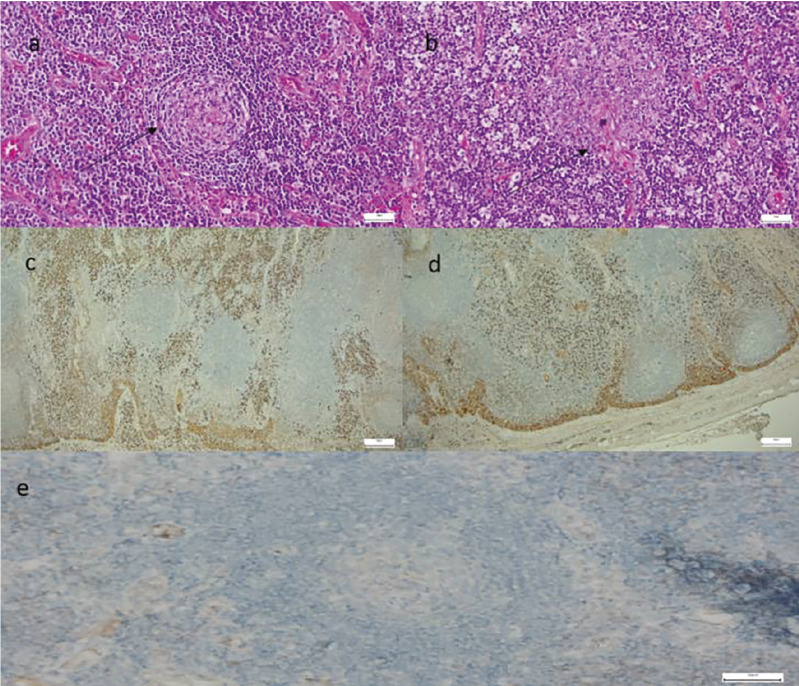
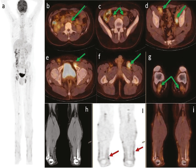
Herein, we describe a rare case of multicentric Castleman disease with multiple lymph node and skin involvement. Ultrasonography of a 38-year-old patient with weakness and fever revealed multiple lymphadenopathies in both inguinal regions. Diagnosed via lymph node biopsy was Castleman's disease, a plasma cell variant. He was diagnosed with prurigo nodularis, lymphocytic vasculitis, and stasis dermatitis in the biopsies of skin lesions located in different regions. 18F-fluorodeoxyglucose positron emission tomography/computed tomography showed multiple hypermetabolic lymph nodes in the axilla, abdomen, pelvis, and both popliteal areas, multiple hypermetabolic skin thickenings, and skin lesions in both arms, legs, and feet.
期刊介绍:
Molecular Imaging and Radionuclide Therapy (Mol Imaging Radionucl Ther, MIRT) is publishes original research articles, invited reviews, editorials, short communications, letters, consensus statements, guidelines and case reports with a literature review on the topic, in the field of molecular imaging, multimodality imaging, nuclear medicine, radionuclide therapy, radiopharmacy, medical physics, dosimetry and radiobiology.

 求助内容:
求助内容: 应助结果提醒方式:


