Rosa Buonamassa, Giuseppe Addabbo, Francesco Pignatelli, Alfredo Niro, Fedele Passidomo
{"title":"联合跨上皮PRK和PTK治疗棘阿米巴角膜炎后角膜混浊1例。","authors":"Rosa Buonamassa, Giuseppe Addabbo, Francesco Pignatelli, Alfredo Niro, Fedele Passidomo","doi":"10.2147/IMCRJ.S495729","DOIUrl":null,"url":null,"abstract":"<p><strong>Purpose: </strong>To report a case of Corneal Opacity following Acanthamoeba Keratitis (AK) treated successfully with transepithelial customized Photorefractive Keratectomy (PRK) combined with Photorefractive Keratectomy (PRK).</p><p><strong>Methods: </strong>One case report.</p><p><strong>Results: </strong>A 27-year-old woman was referred to our clinic for Acanthamoeba keratitis in her left eye. After 1 year from the infection, the patient returned to our attention for developing a central corneal scar and decreased corrected distance visual acuity (CDVA) in the left eye. The slit-lamp examination showed a central corneal opacity involving anterior stroma. A single-step topography-guided trans-epithelial PRK combined with PTK (CIPTA<sup>®</sup>2 software, iVis Technologies) was performed in the left eye. After surface ablation using PRK, PTK was performed using masking agents (1% hydroxymethylcellulose) to smooth the ablated surface. Subsequently, 0.02% Mitomycin C was applied over the ablated surface. At the 1-month follow-up, a resolution of the corneal opacities was observed, with a visual improvement to 20/20, which was maintained at the 3-, 6-, and 12-months follow-up. Furthermore, there was an improvement in spherical equivalent and corneal morphological irregularity index.</p><p><strong>Conclusion: </strong>Corneal opacity following AK may be successfully treated using a combined topography-guided trans-epithelial PRK and PTK in selected patients.</p>","PeriodicalId":14337,"journal":{"name":"International Medical Case Reports Journal","volume":"18 ","pages":"629-635"},"PeriodicalIF":0.7000,"publicationDate":"2025-05-28","publicationTypes":"Journal Article","fieldsOfStudy":null,"isOpenAccess":false,"openAccessPdf":"https://www.ncbi.nlm.nih.gov/pmc/articles/PMC12127525/pdf/","citationCount":"0","resultStr":"{\"title\":\"Combined Trans-Epithelial PRK and PTK for Treatment of Corneal Opacity Following Acanthamoeba Keratitis: A Case Report.\",\"authors\":\"Rosa Buonamassa, Giuseppe Addabbo, Francesco Pignatelli, Alfredo Niro, Fedele Passidomo\",\"doi\":\"10.2147/IMCRJ.S495729\",\"DOIUrl\":null,\"url\":null,\"abstract\":\"<p><strong>Purpose: </strong>To report a case of Corneal Opacity following Acanthamoeba Keratitis (AK) treated successfully with transepithelial customized Photorefractive Keratectomy (PRK) combined with Photorefractive Keratectomy (PRK).</p><p><strong>Methods: </strong>One case report.</p><p><strong>Results: </strong>A 27-year-old woman was referred to our clinic for Acanthamoeba keratitis in her left eye. After 1 year from the infection, the patient returned to our attention for developing a central corneal scar and decreased corrected distance visual acuity (CDVA) in the left eye. The slit-lamp examination showed a central corneal opacity involving anterior stroma. A single-step topography-guided trans-epithelial PRK combined with PTK (CIPTA<sup>®</sup>2 software, iVis Technologies) was performed in the left eye. After surface ablation using PRK, PTK was performed using masking agents (1% hydroxymethylcellulose) to smooth the ablated surface. Subsequently, 0.02% Mitomycin C was applied over the ablated surface. At the 1-month follow-up, a resolution of the corneal opacities was observed, with a visual improvement to 20/20, which was maintained at the 3-, 6-, and 12-months follow-up. Furthermore, there was an improvement in spherical equivalent and corneal morphological irregularity index.</p><p><strong>Conclusion: </strong>Corneal opacity following AK may be successfully treated using a combined topography-guided trans-epithelial PRK and PTK in selected patients.</p>\",\"PeriodicalId\":14337,\"journal\":{\"name\":\"International Medical Case Reports Journal\",\"volume\":\"18 \",\"pages\":\"629-635\"},\"PeriodicalIF\":0.7000,\"publicationDate\":\"2025-05-28\",\"publicationTypes\":\"Journal Article\",\"fieldsOfStudy\":null,\"isOpenAccess\":false,\"openAccessPdf\":\"https://www.ncbi.nlm.nih.gov/pmc/articles/PMC12127525/pdf/\",\"citationCount\":\"0\",\"resultStr\":null,\"platform\":\"Semanticscholar\",\"paperid\":null,\"PeriodicalName\":\"International Medical Case Reports Journal\",\"FirstCategoryId\":\"1085\",\"ListUrlMain\":\"https://doi.org/10.2147/IMCRJ.S495729\",\"RegionNum\":0,\"RegionCategory\":null,\"ArticlePicture\":[],\"TitleCN\":null,\"AbstractTextCN\":null,\"PMCID\":null,\"EPubDate\":\"2025/1/1 0:00:00\",\"PubModel\":\"eCollection\",\"JCR\":\"Q3\",\"JCRName\":\"MEDICINE, GENERAL & INTERNAL\",\"Score\":null,\"Total\":0}","platform":"Semanticscholar","paperid":null,"PeriodicalName":"International Medical Case Reports Journal","FirstCategoryId":"1085","ListUrlMain":"https://doi.org/10.2147/IMCRJ.S495729","RegionNum":0,"RegionCategory":null,"ArticlePicture":[],"TitleCN":null,"AbstractTextCN":null,"PMCID":null,"EPubDate":"2025/1/1 0:00:00","PubModel":"eCollection","JCR":"Q3","JCRName":"MEDICINE, GENERAL & INTERNAL","Score":null,"Total":0}
Combined Trans-Epithelial PRK and PTK for Treatment of Corneal Opacity Following Acanthamoeba Keratitis: A Case Report.
Purpose: To report a case of Corneal Opacity following Acanthamoeba Keratitis (AK) treated successfully with transepithelial customized Photorefractive Keratectomy (PRK) combined with Photorefractive Keratectomy (PRK).
Methods: One case report.
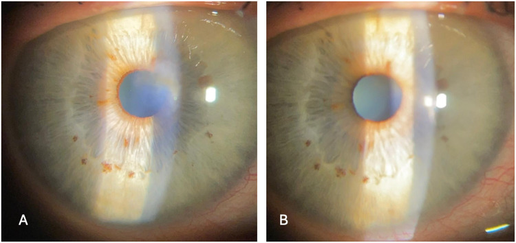
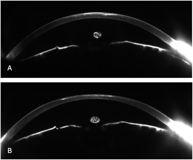
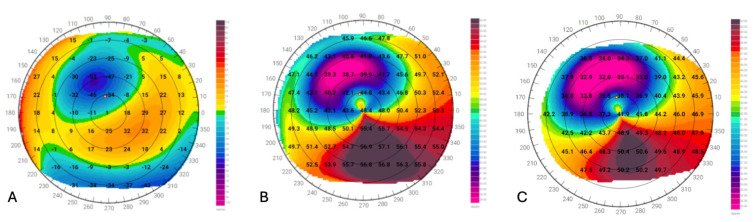
Results: A 27-year-old woman was referred to our clinic for Acanthamoeba keratitis in her left eye. After 1 year from the infection, the patient returned to our attention for developing a central corneal scar and decreased corrected distance visual acuity (CDVA) in the left eye. The slit-lamp examination showed a central corneal opacity involving anterior stroma. A single-step topography-guided trans-epithelial PRK combined with PTK (CIPTA®2 software, iVis Technologies) was performed in the left eye. After surface ablation using PRK, PTK was performed using masking agents (1% hydroxymethylcellulose) to smooth the ablated surface. Subsequently, 0.02% Mitomycin C was applied over the ablated surface. At the 1-month follow-up, a resolution of the corneal opacities was observed, with a visual improvement to 20/20, which was maintained at the 3-, 6-, and 12-months follow-up. Furthermore, there was an improvement in spherical equivalent and corneal morphological irregularity index.
Conclusion: Corneal opacity following AK may be successfully treated using a combined topography-guided trans-epithelial PRK and PTK in selected patients.
期刊介绍:
International Medical Case Reports Journal is an international, peer-reviewed, open access, online journal publishing original case reports from all medical specialties. Submissions should not normally exceed 3,000 words or 4 published pages including figures, diagrams and references. As of 1st April 2019, the International Medical Case Reports Journal will no longer consider meta-analyses for publication.

 求助内容:
求助内容: 应助结果提醒方式:


