Yafei Zhang, Mimi Xu, Yu Wang, Fang Yu, Xinxin Chen, Guangfa Wang, Kui Zhao, Hong Yang, Xinhui Su
{"title":"18F-FAPI-04 PET/CT在腹膜转移患者中的特点及预测治疗效果,与18F-FDG PET/CT进行头对头比较。","authors":"Yafei Zhang, Mimi Xu, Yu Wang, Fang Yu, Xinxin Chen, Guangfa Wang, Kui Zhao, Hong Yang, Xinhui Su","doi":"10.1186/s40644-025-00887-9","DOIUrl":null,"url":null,"abstract":"<p><strong>Background: </strong><sup>18</sup>F-FAPI-04 PET/CT shows promise in detecting peritoneal metastases (PM), but its superiority over <sup>18</sup>F-FDG PET/CT for lesion detection and predicting chemotherapy benefit remains unclear.</p><p><strong>Purpose: </strong>To compare <sup>18</sup>F-FAPI-04 and <sup>18</sup>F-FDG PET/CT imaging features in PM and assess predictive value of <sup>18</sup>F-FAPI-04 for chemotherapy efficacy.</p><p><strong>Methods: </strong>39 pathologically confirmed PM patients with digestive malignancies underwent concurrent <sup>18</sup>F-FAPI-04 and <sup>18</sup>F-FDG PET/CT. Semi-quantitative parameters, including SUV<sub>max</sub>, tumor/liver ratio (T/L), tumor/mediastinal blood pool ratio (T/B), were analyzed. The tracer uptake was compared via Wilcoxon tests. The relationships between <sup>18</sup>F-FAPI-04 uptake with FAP and α-SMA expression were analyzed using Pearson correlation. Patients were divided into different short-term outcome groups (responders vs. non-responders) according to RECIST criteria (v.1.1) after chemotherapy. Post-chemotherapy outcomes were evaluated using logistic regression.</p><p><strong>Results: </strong>Patients (median age 62; 16 females, 23 males) included pancreatic (n = 17), cholangiocarcinoma (n = 8), gastric (n = 6), and colorectal cancers (n = 8). <sup>18</sup>F-FAPI-04 demonstrated significantly higher SUV<sub>max</sub>, T/L, and T/B than <sup>18</sup>F-FDG (P < 0.05). Pancreaticobiliary cancers (pancreatic/cholangiocarcinoma) exhibited higher 18F-FAPI-04 uptake than gastroenteric cancers (gastric/colorectal) (P < 0.05), though no differences existed within subgroups. <sup>18</sup>F-FAPI-04 parameters positively correlated with FAP and α-SMA expression. In univariate analysis, <sup>18</sup>F-FAPI-04 uptake differed significantly between responders and non-responders. Multivariate analysis identified SUV<sub>max</sub> as an independent predictor (OR = 1.354, 95%CI:1.025-1.788, P = 0.033). Optimal <sup>18</sup>F-FAPI-04 cut-offs for distinguishing outcomes were SUV<sub>max</sub>=11.05 (AUC = 0.783; sensitivity = 70.60%, specificity = 80.40%), T/L = 7.53 (AUC = 0.717; 58.82%, 81.82%), and T/B = 8.76 (AUC = 0.751; 64.71%, 86.37%).</p><p><strong>Conclusion: </strong><sup>18</sup>F-FAPI-04 PET/CT outperforms <sup>18</sup>F-FDG in PM detection, with semi-quantitative parameters predicting chemotherapy response.</p>","PeriodicalId":9548,"journal":{"name":"Cancer Imaging","volume":"25 1","pages":"66"},"PeriodicalIF":3.5000,"publicationDate":"2025-06-02","publicationTypes":"Journal Article","fieldsOfStudy":null,"isOpenAccess":false,"openAccessPdf":"https://www.ncbi.nlm.nih.gov/pmc/articles/PMC12128257/pdf/","citationCount":"0","resultStr":"{\"title\":\"Characteristics of <sup>18</sup>F-FAPI-04 PET/CT in patients with peritoneal metastasis and to predict treatment efficacy, a head-to-head comparison with <sup>18</sup>F-FDG PET/CT.\",\"authors\":\"Yafei Zhang, Mimi Xu, Yu Wang, Fang Yu, Xinxin Chen, Guangfa Wang, Kui Zhao, Hong Yang, Xinhui Su\",\"doi\":\"10.1186/s40644-025-00887-9\",\"DOIUrl\":null,\"url\":null,\"abstract\":\"<p><strong>Background: </strong><sup>18</sup>F-FAPI-04 PET/CT shows promise in detecting peritoneal metastases (PM), but its superiority over <sup>18</sup>F-FDG PET/CT for lesion detection and predicting chemotherapy benefit remains unclear.</p><p><strong>Purpose: </strong>To compare <sup>18</sup>F-FAPI-04 and <sup>18</sup>F-FDG PET/CT imaging features in PM and assess predictive value of <sup>18</sup>F-FAPI-04 for chemotherapy efficacy.</p><p><strong>Methods: </strong>39 pathologically confirmed PM patients with digestive malignancies underwent concurrent <sup>18</sup>F-FAPI-04 and <sup>18</sup>F-FDG PET/CT. Semi-quantitative parameters, including SUV<sub>max</sub>, tumor/liver ratio (T/L), tumor/mediastinal blood pool ratio (T/B), were analyzed. The tracer uptake was compared via Wilcoxon tests. The relationships between <sup>18</sup>F-FAPI-04 uptake with FAP and α-SMA expression were analyzed using Pearson correlation. Patients were divided into different short-term outcome groups (responders vs. non-responders) according to RECIST criteria (v.1.1) after chemotherapy. Post-chemotherapy outcomes were evaluated using logistic regression.</p><p><strong>Results: </strong>Patients (median age 62; 16 females, 23 males) included pancreatic (n = 17), cholangiocarcinoma (n = 8), gastric (n = 6), and colorectal cancers (n = 8). <sup>18</sup>F-FAPI-04 demonstrated significantly higher SUV<sub>max</sub>, T/L, and T/B than <sup>18</sup>F-FDG (P < 0.05). Pancreaticobiliary cancers (pancreatic/cholangiocarcinoma) exhibited higher 18F-FAPI-04 uptake than gastroenteric cancers (gastric/colorectal) (P < 0.05), though no differences existed within subgroups. <sup>18</sup>F-FAPI-04 parameters positively correlated with FAP and α-SMA expression. In univariate analysis, <sup>18</sup>F-FAPI-04 uptake differed significantly between responders and non-responders. Multivariate analysis identified SUV<sub>max</sub> as an independent predictor (OR = 1.354, 95%CI:1.025-1.788, P = 0.033). Optimal <sup>18</sup>F-FAPI-04 cut-offs for distinguishing outcomes were SUV<sub>max</sub>=11.05 (AUC = 0.783; sensitivity = 70.60%, specificity = 80.40%), T/L = 7.53 (AUC = 0.717; 58.82%, 81.82%), and T/B = 8.76 (AUC = 0.751; 64.71%, 86.37%).</p><p><strong>Conclusion: </strong><sup>18</sup>F-FAPI-04 PET/CT outperforms <sup>18</sup>F-FDG in PM detection, with semi-quantitative parameters predicting chemotherapy response.</p>\",\"PeriodicalId\":9548,\"journal\":{\"name\":\"Cancer Imaging\",\"volume\":\"25 1\",\"pages\":\"66\"},\"PeriodicalIF\":3.5000,\"publicationDate\":\"2025-06-02\",\"publicationTypes\":\"Journal Article\",\"fieldsOfStudy\":null,\"isOpenAccess\":false,\"openAccessPdf\":\"https://www.ncbi.nlm.nih.gov/pmc/articles/PMC12128257/pdf/\",\"citationCount\":\"0\",\"resultStr\":null,\"platform\":\"Semanticscholar\",\"paperid\":null,\"PeriodicalName\":\"Cancer Imaging\",\"FirstCategoryId\":\"3\",\"ListUrlMain\":\"https://doi.org/10.1186/s40644-025-00887-9\",\"RegionNum\":2,\"RegionCategory\":\"医学\",\"ArticlePicture\":[],\"TitleCN\":null,\"AbstractTextCN\":null,\"PMCID\":null,\"EPubDate\":\"\",\"PubModel\":\"\",\"JCR\":\"Q2\",\"JCRName\":\"ONCOLOGY\",\"Score\":null,\"Total\":0}","platform":"Semanticscholar","paperid":null,"PeriodicalName":"Cancer Imaging","FirstCategoryId":"3","ListUrlMain":"https://doi.org/10.1186/s40644-025-00887-9","RegionNum":2,"RegionCategory":"医学","ArticlePicture":[],"TitleCN":null,"AbstractTextCN":null,"PMCID":null,"EPubDate":"","PubModel":"","JCR":"Q2","JCRName":"ONCOLOGY","Score":null,"Total":0}
Characteristics of 18F-FAPI-04 PET/CT in patients with peritoneal metastasis and to predict treatment efficacy, a head-to-head comparison with 18F-FDG PET/CT.
Background: 18F-FAPI-04 PET/CT shows promise in detecting peritoneal metastases (PM), but its superiority over 18F-FDG PET/CT for lesion detection and predicting chemotherapy benefit remains unclear.
Purpose: To compare 18F-FAPI-04 and 18F-FDG PET/CT imaging features in PM and assess predictive value of 18F-FAPI-04 for chemotherapy efficacy.
Methods: 39 pathologically confirmed PM patients with digestive malignancies underwent concurrent 18F-FAPI-04 and 18F-FDG PET/CT. Semi-quantitative parameters, including SUVmax, tumor/liver ratio (T/L), tumor/mediastinal blood pool ratio (T/B), were analyzed. The tracer uptake was compared via Wilcoxon tests. The relationships between 18F-FAPI-04 uptake with FAP and α-SMA expression were analyzed using Pearson correlation. Patients were divided into different short-term outcome groups (responders vs. non-responders) according to RECIST criteria (v.1.1) after chemotherapy. Post-chemotherapy outcomes were evaluated using logistic regression.
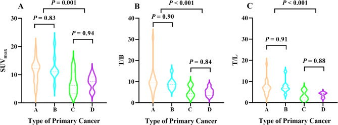
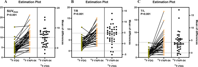
Results: Patients (median age 62; 16 females, 23 males) included pancreatic (n = 17), cholangiocarcinoma (n = 8), gastric (n = 6), and colorectal cancers (n = 8). 18F-FAPI-04 demonstrated significantly higher SUVmax, T/L, and T/B than 18F-FDG (P < 0.05). Pancreaticobiliary cancers (pancreatic/cholangiocarcinoma) exhibited higher 18F-FAPI-04 uptake than gastroenteric cancers (gastric/colorectal) (P < 0.05), though no differences existed within subgroups. 18F-FAPI-04 parameters positively correlated with FAP and α-SMA expression. In univariate analysis, 18F-FAPI-04 uptake differed significantly between responders and non-responders. Multivariate analysis identified SUVmax as an independent predictor (OR = 1.354, 95%CI:1.025-1.788, P = 0.033). Optimal 18F-FAPI-04 cut-offs for distinguishing outcomes were SUVmax=11.05 (AUC = 0.783; sensitivity = 70.60%, specificity = 80.40%), T/L = 7.53 (AUC = 0.717; 58.82%, 81.82%), and T/B = 8.76 (AUC = 0.751; 64.71%, 86.37%).
Conclusion: 18F-FAPI-04 PET/CT outperforms 18F-FDG in PM detection, with semi-quantitative parameters predicting chemotherapy response.
Cancer ImagingONCOLOGY-RADIOLOGY, NUCLEAR MEDICINE & MEDICAL IMAGING
CiteScore
7.00
自引率
0.00%
发文量
66
审稿时长
>12 weeks
期刊介绍:
Cancer Imaging is an open access, peer-reviewed journal publishing original articles, reviews and editorials written by expert international radiologists working in oncology.
The journal encompasses CT, MR, PET, ultrasound, radionuclide and multimodal imaging in all kinds of malignant tumours, plus new developments, techniques and innovations. Topics of interest include:
Breast Imaging
Chest
Complications of treatment
Ear, Nose & Throat
Gastrointestinal
Hepatobiliary & Pancreatic
Imaging biomarkers
Interventional
Lymphoma
Measurement of tumour response
Molecular functional imaging
Musculoskeletal
Neuro oncology
Nuclear Medicine
Paediatric.


 求助内容:
求助内容: 应助结果提醒方式:


