{"title":"犬中央分割肝切除术后的进展性肝病。","authors":"SJ Wood, GL Hosgood, F Coiacetto","doi":"10.1111/avj.13452","DOIUrl":null,"url":null,"abstract":"<p>The consequences of large-volume hepatectomy on the remaining liver in the dog are not documented. This case report documents a progressive hepatopathy after central division hepatectomy in a 10-year-old female Lhasa Apso dog. Serum biochemistry at presentation for chronic urinary incontinence indicated a hepatopathy, with a liver mass identified on subsequent ultrasound. Referral for computed tomography (CT) (day 76) demonstrated a central division liver mass (~204cm<sup>3</sup>). A central division hepatectomy (right medial and quadrate lobes; gallbladder) was performed, with microscopic examination confirming a hepatocellular carcinoma. Serial biochemistry over a three-and-a-half-year follow-up documented progressive hepatopathy. Serial CT hepatic volumetry (day 76, 136, 1041) documented gross enlargement of the residual right lateral liver lobe and gross reduction of the residual left medial and lateral liver lobes. Biopsies of the left and right liver (day 187) demonstrated disparate pathology with microscopic features consistent with regeneration in the right liver and elevated portal vein pressure (PVP) in the left liver. This report documents progressive hepatopathy evidenced by serum biochemistry, disparate and persistent gross liver changes on posthepatectomy CT, and microscopic changes consistent with elevated PVP. Findings from this case demonstrate similarities to posthepatectomy liver failure (PHLF) and small-for-size-and-flow syndrome (SFSS) as reported in people and animal models. The progressive hepatopathy posthepatectomy in this case prompts consideration and further investigation for the development of PHLF and SFSS in the dog.</p>","PeriodicalId":8661,"journal":{"name":"Australian Veterinary Journal","volume":"103 7","pages":"422-429"},"PeriodicalIF":1.7000,"publicationDate":"2025-06-02","publicationTypes":"Journal Article","fieldsOfStudy":null,"isOpenAccess":false,"openAccessPdf":"https://onlinelibrary.wiley.com/doi/epdf/10.1111/avj.13452","citationCount":"0","resultStr":"{\"title\":\"Progressive hepatopathy after central division hepatectomy in a dog\",\"authors\":\"SJ Wood, GL Hosgood, F Coiacetto\",\"doi\":\"10.1111/avj.13452\",\"DOIUrl\":null,\"url\":null,\"abstract\":\"<p>The consequences of large-volume hepatectomy on the remaining liver in the dog are not documented. This case report documents a progressive hepatopathy after central division hepatectomy in a 10-year-old female Lhasa Apso dog. Serum biochemistry at presentation for chronic urinary incontinence indicated a hepatopathy, with a liver mass identified on subsequent ultrasound. Referral for computed tomography (CT) (day 76) demonstrated a central division liver mass (~204cm<sup>3</sup>). A central division hepatectomy (right medial and quadrate lobes; gallbladder) was performed, with microscopic examination confirming a hepatocellular carcinoma. Serial biochemistry over a three-and-a-half-year follow-up documented progressive hepatopathy. Serial CT hepatic volumetry (day 76, 136, 1041) documented gross enlargement of the residual right lateral liver lobe and gross reduction of the residual left medial and lateral liver lobes. Biopsies of the left and right liver (day 187) demonstrated disparate pathology with microscopic features consistent with regeneration in the right liver and elevated portal vein pressure (PVP) in the left liver. This report documents progressive hepatopathy evidenced by serum biochemistry, disparate and persistent gross liver changes on posthepatectomy CT, and microscopic changes consistent with elevated PVP. Findings from this case demonstrate similarities to posthepatectomy liver failure (PHLF) and small-for-size-and-flow syndrome (SFSS) as reported in people and animal models. The progressive hepatopathy posthepatectomy in this case prompts consideration and further investigation for the development of PHLF and SFSS in the dog.</p>\",\"PeriodicalId\":8661,\"journal\":{\"name\":\"Australian Veterinary Journal\",\"volume\":\"103 7\",\"pages\":\"422-429\"},\"PeriodicalIF\":1.7000,\"publicationDate\":\"2025-06-02\",\"publicationTypes\":\"Journal Article\",\"fieldsOfStudy\":null,\"isOpenAccess\":false,\"openAccessPdf\":\"https://onlinelibrary.wiley.com/doi/epdf/10.1111/avj.13452\",\"citationCount\":\"0\",\"resultStr\":null,\"platform\":\"Semanticscholar\",\"paperid\":null,\"PeriodicalName\":\"Australian Veterinary Journal\",\"FirstCategoryId\":\"97\",\"ListUrlMain\":\"https://onlinelibrary.wiley.com/doi/10.1111/avj.13452\",\"RegionNum\":4,\"RegionCategory\":\"农林科学\",\"ArticlePicture\":[],\"TitleCN\":null,\"AbstractTextCN\":null,\"PMCID\":null,\"EPubDate\":\"\",\"PubModel\":\"\",\"JCR\":\"Q2\",\"JCRName\":\"VETERINARY SCIENCES\",\"Score\":null,\"Total\":0}","platform":"Semanticscholar","paperid":null,"PeriodicalName":"Australian Veterinary Journal","FirstCategoryId":"97","ListUrlMain":"https://onlinelibrary.wiley.com/doi/10.1111/avj.13452","RegionNum":4,"RegionCategory":"农林科学","ArticlePicture":[],"TitleCN":null,"AbstractTextCN":null,"PMCID":null,"EPubDate":"","PubModel":"","JCR":"Q2","JCRName":"VETERINARY SCIENCES","Score":null,"Total":0}
Progressive hepatopathy after central division hepatectomy in a dog
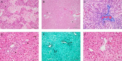
The consequences of large-volume hepatectomy on the remaining liver in the dog are not documented. This case report documents a progressive hepatopathy after central division hepatectomy in a 10-year-old female Lhasa Apso dog. Serum biochemistry at presentation for chronic urinary incontinence indicated a hepatopathy, with a liver mass identified on subsequent ultrasound. Referral for computed tomography (CT) (day 76) demonstrated a central division liver mass (~204cm3). A central division hepatectomy (right medial and quadrate lobes; gallbladder) was performed, with microscopic examination confirming a hepatocellular carcinoma. Serial biochemistry over a three-and-a-half-year follow-up documented progressive hepatopathy. Serial CT hepatic volumetry (day 76, 136, 1041) documented gross enlargement of the residual right lateral liver lobe and gross reduction of the residual left medial and lateral liver lobes. Biopsies of the left and right liver (day 187) demonstrated disparate pathology with microscopic features consistent with regeneration in the right liver and elevated portal vein pressure (PVP) in the left liver. This report documents progressive hepatopathy evidenced by serum biochemistry, disparate and persistent gross liver changes on posthepatectomy CT, and microscopic changes consistent with elevated PVP. Findings from this case demonstrate similarities to posthepatectomy liver failure (PHLF) and small-for-size-and-flow syndrome (SFSS) as reported in people and animal models. The progressive hepatopathy posthepatectomy in this case prompts consideration and further investigation for the development of PHLF and SFSS in the dog.
期刊介绍:
Over the past 80 years, the Australian Veterinary Journal (AVJ) has been providing the veterinary profession with leading edge clinical and scientific research, case reports, reviews. news and timely coverage of industry issues. AJV is Australia''s premier veterinary science text and is distributed monthly to over 5,500 Australian Veterinary Association members and subscribers.
 求助内容:
求助内容: 应助结果提醒方式:


