{"title":"跑步加速度与腰椎间盘的T2磁共振成像值相关。","authors":"Takayoshi Hakkaku, Yoshiaki Kubo, Koji Koyama, Koichi Nakazato, Takashi Okada, Kenji Hiranuma","doi":"10.1155/tsm2/5930823","DOIUrl":null,"url":null,"abstract":"<p><p><b>Background:</b> Running can contribute to both beneficial and detrimental responses in the intervertebral discs (IVDs). To better understand these effects, we investigated the relationship between loading directions during slow running and the rapid changes in T2 times occurring in the lumbar IVDs before and after running. <b>Methods:</b> Sixteen healthy male students were fitted with a triaxial accelerator and ran on a treadmill at 8 km/h for 1 minute. Three lumbar T2 times from the L3/L4 to L5/S1 levels were measured before, immediately after, and 30 min postexercise via magnetic resonance imaging (MRI). The analysis focused on five regions of interest within each disc. <b>Results:</b> Acceleration was 0.23 ± 0.06 root mean square in the mediolateral (<i>X</i>-axis), 1.37 ± 0.08 in the vertical (<i>Y</i>-axis), and 0.30 ± 0.06 in the anteroposterior (<i>Z</i>-axis) direction. A strong correlation was observed between the T2 relaxation times and acceleration, particularly in the <i>Z</i>-axis. At L3/L4, a positive correlation was observed for the posterior nucleus (<i>r</i> = 0.72, <i>p</i>=0.002, <i>R</i> <sup>2</sup> = 0.59). At L4/L5, a positive correlation was observed for the central nucleus (<i>r</i> = 0.73, <i>p</i>=0.003, <i>R</i> <sup>2</sup> = 0.49). At L5/S1, a negative correlation was observed for the anterior annulus fibrosus (<i>r</i> = -0.73, <i>p</i>=0.01, <i>R</i> <sup>2</sup> = 0.48). <b>Conclusion:</b> These results suggest that anteroposterior loading may play a significant role in the response of the IVDs to running.</p>","PeriodicalId":75247,"journal":{"name":"Translational sports medicine","volume":"2025 ","pages":"5930823"},"PeriodicalIF":1.9000,"publicationDate":"2025-05-24","publicationTypes":"Journal Article","fieldsOfStudy":null,"isOpenAccess":false,"openAccessPdf":"https://www.ncbi.nlm.nih.gov/pmc/articles/PMC12126259/pdf/","citationCount":"0","resultStr":"{\"title\":\"Running Acceleration Correlates With T2 Magnetic Resonance Imaging Values of the Lumber Intervertebral Disc.\",\"authors\":\"Takayoshi Hakkaku, Yoshiaki Kubo, Koji Koyama, Koichi Nakazato, Takashi Okada, Kenji Hiranuma\",\"doi\":\"10.1155/tsm2/5930823\",\"DOIUrl\":null,\"url\":null,\"abstract\":\"<p><p><b>Background:</b> Running can contribute to both beneficial and detrimental responses in the intervertebral discs (IVDs). To better understand these effects, we investigated the relationship between loading directions during slow running and the rapid changes in T2 times occurring in the lumbar IVDs before and after running. <b>Methods:</b> Sixteen healthy male students were fitted with a triaxial accelerator and ran on a treadmill at 8 km/h for 1 minute. Three lumbar T2 times from the L3/L4 to L5/S1 levels were measured before, immediately after, and 30 min postexercise via magnetic resonance imaging (MRI). The analysis focused on five regions of interest within each disc. <b>Results:</b> Acceleration was 0.23 ± 0.06 root mean square in the mediolateral (<i>X</i>-axis), 1.37 ± 0.08 in the vertical (<i>Y</i>-axis), and 0.30 ± 0.06 in the anteroposterior (<i>Z</i>-axis) direction. A strong correlation was observed between the T2 relaxation times and acceleration, particularly in the <i>Z</i>-axis. At L3/L4, a positive correlation was observed for the posterior nucleus (<i>r</i> = 0.72, <i>p</i>=0.002, <i>R</i> <sup>2</sup> = 0.59). At L4/L5, a positive correlation was observed for the central nucleus (<i>r</i> = 0.73, <i>p</i>=0.003, <i>R</i> <sup>2</sup> = 0.49). At L5/S1, a negative correlation was observed for the anterior annulus fibrosus (<i>r</i> = -0.73, <i>p</i>=0.01, <i>R</i> <sup>2</sup> = 0.48). <b>Conclusion:</b> These results suggest that anteroposterior loading may play a significant role in the response of the IVDs to running.</p>\",\"PeriodicalId\":75247,\"journal\":{\"name\":\"Translational sports medicine\",\"volume\":\"2025 \",\"pages\":\"5930823\"},\"PeriodicalIF\":1.9000,\"publicationDate\":\"2025-05-24\",\"publicationTypes\":\"Journal Article\",\"fieldsOfStudy\":null,\"isOpenAccess\":false,\"openAccessPdf\":\"https://www.ncbi.nlm.nih.gov/pmc/articles/PMC12126259/pdf/\",\"citationCount\":\"0\",\"resultStr\":null,\"platform\":\"Semanticscholar\",\"paperid\":null,\"PeriodicalName\":\"Translational sports medicine\",\"FirstCategoryId\":\"1085\",\"ListUrlMain\":\"https://doi.org/10.1155/tsm2/5930823\",\"RegionNum\":0,\"RegionCategory\":null,\"ArticlePicture\":[],\"TitleCN\":null,\"AbstractTextCN\":null,\"PMCID\":null,\"EPubDate\":\"2025/1/1 0:00:00\",\"PubModel\":\"eCollection\",\"JCR\":\"Q3\",\"JCRName\":\"SPORT SCIENCES\",\"Score\":null,\"Total\":0}","platform":"Semanticscholar","paperid":null,"PeriodicalName":"Translational sports medicine","FirstCategoryId":"1085","ListUrlMain":"https://doi.org/10.1155/tsm2/5930823","RegionNum":0,"RegionCategory":null,"ArticlePicture":[],"TitleCN":null,"AbstractTextCN":null,"PMCID":null,"EPubDate":"2025/1/1 0:00:00","PubModel":"eCollection","JCR":"Q3","JCRName":"SPORT SCIENCES","Score":null,"Total":0}
Running Acceleration Correlates With T2 Magnetic Resonance Imaging Values of the Lumber Intervertebral Disc.
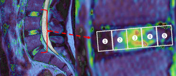
Background: Running can contribute to both beneficial and detrimental responses in the intervertebral discs (IVDs). To better understand these effects, we investigated the relationship between loading directions during slow running and the rapid changes in T2 times occurring in the lumbar IVDs before and after running. Methods: Sixteen healthy male students were fitted with a triaxial accelerator and ran on a treadmill at 8 km/h for 1 minute. Three lumbar T2 times from the L3/L4 to L5/S1 levels were measured before, immediately after, and 30 min postexercise via magnetic resonance imaging (MRI). The analysis focused on five regions of interest within each disc. Results: Acceleration was 0.23 ± 0.06 root mean square in the mediolateral (X-axis), 1.37 ± 0.08 in the vertical (Y-axis), and 0.30 ± 0.06 in the anteroposterior (Z-axis) direction. A strong correlation was observed between the T2 relaxation times and acceleration, particularly in the Z-axis. At L3/L4, a positive correlation was observed for the posterior nucleus (r = 0.72, p=0.002, R2 = 0.59). At L4/L5, a positive correlation was observed for the central nucleus (r = 0.73, p=0.003, R2 = 0.49). At L5/S1, a negative correlation was observed for the anterior annulus fibrosus (r = -0.73, p=0.01, R2 = 0.48). Conclusion: These results suggest that anteroposterior loading may play a significant role in the response of the IVDs to running.

 求助内容:
求助内容: 应助结果提醒方式:


