Moosa Mahmoudi, Kianoosh Mobasseri, Sara Samiei, Ali Labafchi
{"title":"下颌冠状巨骨软骨瘤表现为冠状增生1例罕见临床报告。","authors":"Moosa Mahmoudi, Kianoosh Mobasseri, Sara Samiei, Ali Labafchi","doi":"10.61186/wjps.14.1.105","DOIUrl":null,"url":null,"abstract":"<p><p>Osteochondroma, the most common benign tumor in the axial and appendicular skeleton, presents a unique challenge when encountered in the craniofacial region. We report a rare case of a 35-year-old female with a massive osteochondroma located on the mandibular coronoid process, resulting in a 20-year history of progressive mouth opening limitation, facial asymmetry, and zygomatic deformity. Comprehensive diagnostic procedures, including panoramic radiography and computed tomography (CT) imaging, were employed to accurately assess the extent of the lesion. This case highlights the importance of CT scans and three-dimensional reconstructions in confirming the diagnosis, particularly when panoramic radiographs exhibit limitations. The patient underwent a successful extraoral approach for coronoidectomy and excision of the osteochondroma, addressing both the functional and cosmetic aspects of the condition. Postoperative evaluations revealed significant improvement in mouth opening capacity, with no signs of recurrence during a 12-month follow-up period.</p>","PeriodicalId":23736,"journal":{"name":"World Journal of Plastic Surgery","volume":"14 1","pages":"105-111"},"PeriodicalIF":1.0000,"publicationDate":"2025-01-01","publicationTypes":"Journal Article","fieldsOfStudy":null,"isOpenAccess":false,"openAccessPdf":"https://www.ncbi.nlm.nih.gov/pmc/articles/PMC12124271/pdf/","citationCount":"0","resultStr":"{\"title\":\"A Giant Osteochondroma of Mandibular Coronoid that Mimicking Coronoid Hyperplasia: A Rare Clinical Case Report.\",\"authors\":\"Moosa Mahmoudi, Kianoosh Mobasseri, Sara Samiei, Ali Labafchi\",\"doi\":\"10.61186/wjps.14.1.105\",\"DOIUrl\":null,\"url\":null,\"abstract\":\"<p><p>Osteochondroma, the most common benign tumor in the axial and appendicular skeleton, presents a unique challenge when encountered in the craniofacial region. We report a rare case of a 35-year-old female with a massive osteochondroma located on the mandibular coronoid process, resulting in a 20-year history of progressive mouth opening limitation, facial asymmetry, and zygomatic deformity. Comprehensive diagnostic procedures, including panoramic radiography and computed tomography (CT) imaging, were employed to accurately assess the extent of the lesion. This case highlights the importance of CT scans and three-dimensional reconstructions in confirming the diagnosis, particularly when panoramic radiographs exhibit limitations. The patient underwent a successful extraoral approach for coronoidectomy and excision of the osteochondroma, addressing both the functional and cosmetic aspects of the condition. Postoperative evaluations revealed significant improvement in mouth opening capacity, with no signs of recurrence during a 12-month follow-up period.</p>\",\"PeriodicalId\":23736,\"journal\":{\"name\":\"World Journal of Plastic Surgery\",\"volume\":\"14 1\",\"pages\":\"105-111\"},\"PeriodicalIF\":1.0000,\"publicationDate\":\"2025-01-01\",\"publicationTypes\":\"Journal Article\",\"fieldsOfStudy\":null,\"isOpenAccess\":false,\"openAccessPdf\":\"https://www.ncbi.nlm.nih.gov/pmc/articles/PMC12124271/pdf/\",\"citationCount\":\"0\",\"resultStr\":null,\"platform\":\"Semanticscholar\",\"paperid\":null,\"PeriodicalName\":\"World Journal of Plastic Surgery\",\"FirstCategoryId\":\"1085\",\"ListUrlMain\":\"https://doi.org/10.61186/wjps.14.1.105\",\"RegionNum\":0,\"RegionCategory\":null,\"ArticlePicture\":[],\"TitleCN\":null,\"AbstractTextCN\":null,\"PMCID\":null,\"EPubDate\":\"\",\"PubModel\":\"\",\"JCR\":\"Q3\",\"JCRName\":\"SURGERY\",\"Score\":null,\"Total\":0}","platform":"Semanticscholar","paperid":null,"PeriodicalName":"World Journal of Plastic Surgery","FirstCategoryId":"1085","ListUrlMain":"https://doi.org/10.61186/wjps.14.1.105","RegionNum":0,"RegionCategory":null,"ArticlePicture":[],"TitleCN":null,"AbstractTextCN":null,"PMCID":null,"EPubDate":"","PubModel":"","JCR":"Q3","JCRName":"SURGERY","Score":null,"Total":0}
A Giant Osteochondroma of Mandibular Coronoid that Mimicking Coronoid Hyperplasia: A Rare Clinical Case Report.
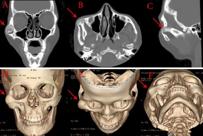
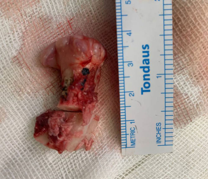
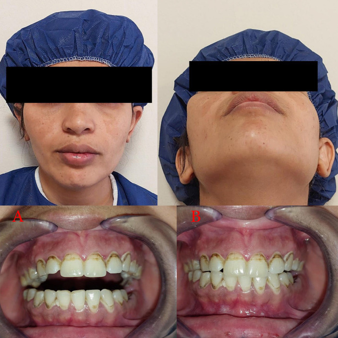
Osteochondroma, the most common benign tumor in the axial and appendicular skeleton, presents a unique challenge when encountered in the craniofacial region. We report a rare case of a 35-year-old female with a massive osteochondroma located on the mandibular coronoid process, resulting in a 20-year history of progressive mouth opening limitation, facial asymmetry, and zygomatic deformity. Comprehensive diagnostic procedures, including panoramic radiography and computed tomography (CT) imaging, were employed to accurately assess the extent of the lesion. This case highlights the importance of CT scans and three-dimensional reconstructions in confirming the diagnosis, particularly when panoramic radiographs exhibit limitations. The patient underwent a successful extraoral approach for coronoidectomy and excision of the osteochondroma, addressing both the functional and cosmetic aspects of the condition. Postoperative evaluations revealed significant improvement in mouth opening capacity, with no signs of recurrence during a 12-month follow-up period.

 求助内容:
求助内容: 应助结果提醒方式:


