Ghada Bouslama, Aya Mtiri, Hajer Zidani, Lamia Oualha, Souha Ben Youssef
{"title":"牙源性囊肿减压的数字化工作流程:定制可移动装置的设计和制造:一例报告。","authors":"Ghada Bouslama, Aya Mtiri, Hajer Zidani, Lamia Oualha, Souha Ben Youssef","doi":"10.1177/11795476251342354","DOIUrl":null,"url":null,"abstract":"<p><p>Dentigerous cysts are the most common type of developmental odontogenic cysts. Multiple devices has been described for decompression. The current case report describes the use of a custom-made decompression appliance, designed through a digital workflow, in managing dentigerous cysts. A 7-year-old male patient with no prior medical history was referred to our oral surgery department due to swelling on the left side of the lower jaw. Upon intraoral examination and cone-beam computed tomography (CBCT), a provisional diagnosis of an inflammatory dentigerous cyst related to the impacted premolar was made. A digital decompression appliance was planned using EXOCAD (Exocad Gmbh, Darmstadt, Germany), and produced using a stereolithography (SLA) 3D printer. The appliance were delivered on the day of the cystostomy after extraction of the deciduous molar (tooth 85). In this report, the advancements in digital design technologies were explored enabling the creation of customized cyst decompression devices. Various stages of the design process were discussed, including 3D modeling, material selection, and the integration of digital workflows in the fabrication process. Additionally, the benefits of using such devices were addressed, including improved patient outcomes, enhanced precision in treatment, and the reduction of surgical complications.</p>","PeriodicalId":10357,"journal":{"name":"Clinical Medicine Insights. Case Reports","volume":"18 ","pages":"11795476251342354"},"PeriodicalIF":0.6000,"publicationDate":"2025-05-28","publicationTypes":"Journal Article","fieldsOfStudy":null,"isOpenAccess":false,"openAccessPdf":"https://www.ncbi.nlm.nih.gov/pmc/articles/PMC12123102/pdf/","citationCount":"0","resultStr":"{\"title\":\"Digital Workflow for Odontogenic Cyst Decompression: Design and Fabrication of a Custom Removable Device: A Case Report.\",\"authors\":\"Ghada Bouslama, Aya Mtiri, Hajer Zidani, Lamia Oualha, Souha Ben Youssef\",\"doi\":\"10.1177/11795476251342354\",\"DOIUrl\":null,\"url\":null,\"abstract\":\"<p><p>Dentigerous cysts are the most common type of developmental odontogenic cysts. Multiple devices has been described for decompression. The current case report describes the use of a custom-made decompression appliance, designed through a digital workflow, in managing dentigerous cysts. A 7-year-old male patient with no prior medical history was referred to our oral surgery department due to swelling on the left side of the lower jaw. Upon intraoral examination and cone-beam computed tomography (CBCT), a provisional diagnosis of an inflammatory dentigerous cyst related to the impacted premolar was made. A digital decompression appliance was planned using EXOCAD (Exocad Gmbh, Darmstadt, Germany), and produced using a stereolithography (SLA) 3D printer. The appliance were delivered on the day of the cystostomy after extraction of the deciduous molar (tooth 85). In this report, the advancements in digital design technologies were explored enabling the creation of customized cyst decompression devices. Various stages of the design process were discussed, including 3D modeling, material selection, and the integration of digital workflows in the fabrication process. Additionally, the benefits of using such devices were addressed, including improved patient outcomes, enhanced precision in treatment, and the reduction of surgical complications.</p>\",\"PeriodicalId\":10357,\"journal\":{\"name\":\"Clinical Medicine Insights. Case Reports\",\"volume\":\"18 \",\"pages\":\"11795476251342354\"},\"PeriodicalIF\":0.6000,\"publicationDate\":\"2025-05-28\",\"publicationTypes\":\"Journal Article\",\"fieldsOfStudy\":null,\"isOpenAccess\":false,\"openAccessPdf\":\"https://www.ncbi.nlm.nih.gov/pmc/articles/PMC12123102/pdf/\",\"citationCount\":\"0\",\"resultStr\":null,\"platform\":\"Semanticscholar\",\"paperid\":null,\"PeriodicalName\":\"Clinical Medicine Insights. Case Reports\",\"FirstCategoryId\":\"1085\",\"ListUrlMain\":\"https://doi.org/10.1177/11795476251342354\",\"RegionNum\":0,\"RegionCategory\":null,\"ArticlePicture\":[],\"TitleCN\":null,\"AbstractTextCN\":null,\"PMCID\":null,\"EPubDate\":\"2025/1/1 0:00:00\",\"PubModel\":\"eCollection\",\"JCR\":\"Q3\",\"JCRName\":\"MEDICINE, GENERAL & INTERNAL\",\"Score\":null,\"Total\":0}","platform":"Semanticscholar","paperid":null,"PeriodicalName":"Clinical Medicine Insights. Case Reports","FirstCategoryId":"1085","ListUrlMain":"https://doi.org/10.1177/11795476251342354","RegionNum":0,"RegionCategory":null,"ArticlePicture":[],"TitleCN":null,"AbstractTextCN":null,"PMCID":null,"EPubDate":"2025/1/1 0:00:00","PubModel":"eCollection","JCR":"Q3","JCRName":"MEDICINE, GENERAL & INTERNAL","Score":null,"Total":0}
Digital Workflow for Odontogenic Cyst Decompression: Design and Fabrication of a Custom Removable Device: A Case Report.
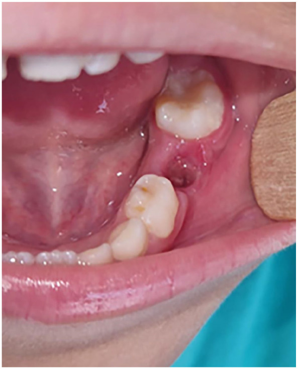
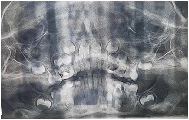
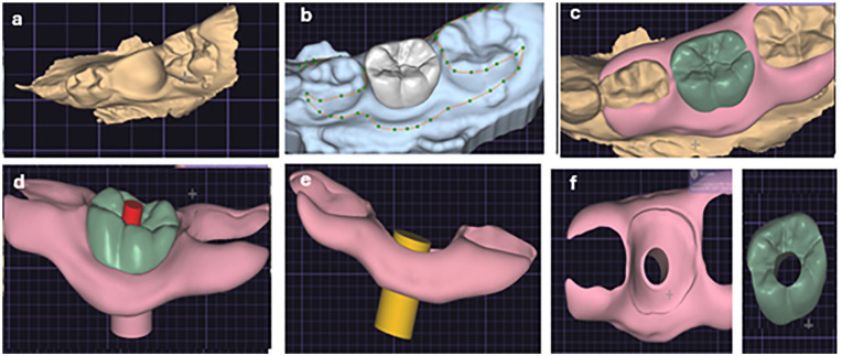
Dentigerous cysts are the most common type of developmental odontogenic cysts. Multiple devices has been described for decompression. The current case report describes the use of a custom-made decompression appliance, designed through a digital workflow, in managing dentigerous cysts. A 7-year-old male patient with no prior medical history was referred to our oral surgery department due to swelling on the left side of the lower jaw. Upon intraoral examination and cone-beam computed tomography (CBCT), a provisional diagnosis of an inflammatory dentigerous cyst related to the impacted premolar was made. A digital decompression appliance was planned using EXOCAD (Exocad Gmbh, Darmstadt, Germany), and produced using a stereolithography (SLA) 3D printer. The appliance were delivered on the day of the cystostomy after extraction of the deciduous molar (tooth 85). In this report, the advancements in digital design technologies were explored enabling the creation of customized cyst decompression devices. Various stages of the design process were discussed, including 3D modeling, material selection, and the integration of digital workflows in the fabrication process. Additionally, the benefits of using such devices were addressed, including improved patient outcomes, enhanced precision in treatment, and the reduction of surgical complications.

 求助内容:
求助内容: 应助结果提醒方式:


