Tim Lyckenvik, Malin Woock, Kalle Johansson, Markus Axelsson, Henrik Zetterberg, Kaj Blennow, Eric Hanse, Pontus Wasling
{"title":"阿片类药物依赖的脑脊液生物标志物:神经免疫激活和离子组成变化的证据,不改变食欲素- a","authors":"Tim Lyckenvik, Malin Woock, Kalle Johansson, Markus Axelsson, Henrik Zetterberg, Kaj Blennow, Eric Hanse, Pontus Wasling","doi":"10.1111/adb.70053","DOIUrl":null,"url":null,"abstract":"<p>Opioid abuse is a severe global health challenge, leading to rising morbidity, mortality, and increasing societal costs. The aim of this study was to investigate neuroinflammation, neuronal damage and potential changes in the orexin system or beta-amyloid metabolism in the cerebrospinal fluid (CSF) of individuals undergoing opioid substitution therapy (OST). This cross-sectional study investigates CSF biomarkers in individuals undergoing OST, compared to control subjects. Participants receiving OST were recruited from the outpatient clinic at the Department of Psychiatry, Sahlgrenska University Hospital, Gothenburg (Sweden). Each participant provided a complete medical history, including details of drug use over the past 6 months, followed by a lumbar puncture to obtain CSF samples. Molecules associated with neuroinflammation, neuronal and glial damage, beta-amyloid metabolism and orexinergic function were analysed in the participants' CSF, alongside electrolyte levels. Specifically, we analysed levels of sTREM-2, YKL-40, IL-1β, IL-6, IL-8, IL-10, TNF-α, AXL, MER, TYRO3, GAS6, NfL, GFAP, total tau (T-tau), phosphorylated tau (P-tau), neurogranin, Aβ40, Aβ42, the Aβ42/Aβ40 ratio, orexin-A, sPDGFR-β and electrolytes. The study included 15 control subjects and 17 in the opioid substitution group. Patients undergoing opioid substitution therapy exhibited elevated levels of sTREM-2, Aβ42/Aβ40 ratio and NfL in their CSF. Conversely, concentrations of Na<sup>+</sup> and Cl<sup>−</sup> were lower compared to controls. No significant differences were found between groups for other biomarkers, including orexin-A. However, when normalized to Aβ40 levels, YKL-40, IL-8, TYRO3 and P-Tau were also elevated in individuals with opioid dependence. Elevated biomarkers of neuroimmune activation, neuronal damage and beta-amyloid metabolism in opioid dependence suggest CNS inflammation as a contributor to its pathophysiology. Reduced electrolyte levels imply disrupted CSF water regulation, possibly linked to impaired glial function. These findings highlight both neural and non-neural mechanisms in opioid dependence.</p>","PeriodicalId":7289,"journal":{"name":"Addiction Biology","volume":"30 6","pages":""},"PeriodicalIF":2.6000,"publicationDate":"2025-06-02","publicationTypes":"Journal Article","fieldsOfStudy":null,"isOpenAccess":false,"openAccessPdf":"https://onlinelibrary.wiley.com/doi/epdf/10.1111/adb.70053","citationCount":"0","resultStr":"{\"title\":\"Cerebrospinal Fluid Biomarkers in Opioid Dependence: Evidence of Neuroimmune Activation and Ion Composition Changes, Without Alteration in Orexin-A\",\"authors\":\"Tim Lyckenvik, Malin Woock, Kalle Johansson, Markus Axelsson, Henrik Zetterberg, Kaj Blennow, Eric Hanse, Pontus Wasling\",\"doi\":\"10.1111/adb.70053\",\"DOIUrl\":null,\"url\":null,\"abstract\":\"<p>Opioid abuse is a severe global health challenge, leading to rising morbidity, mortality, and increasing societal costs. The aim of this study was to investigate neuroinflammation, neuronal damage and potential changes in the orexin system or beta-amyloid metabolism in the cerebrospinal fluid (CSF) of individuals undergoing opioid substitution therapy (OST). This cross-sectional study investigates CSF biomarkers in individuals undergoing OST, compared to control subjects. Participants receiving OST were recruited from the outpatient clinic at the Department of Psychiatry, Sahlgrenska University Hospital, Gothenburg (Sweden). Each participant provided a complete medical history, including details of drug use over the past 6 months, followed by a lumbar puncture to obtain CSF samples. Molecules associated with neuroinflammation, neuronal and glial damage, beta-amyloid metabolism and orexinergic function were analysed in the participants' CSF, alongside electrolyte levels. Specifically, we analysed levels of sTREM-2, YKL-40, IL-1β, IL-6, IL-8, IL-10, TNF-α, AXL, MER, TYRO3, GAS6, NfL, GFAP, total tau (T-tau), phosphorylated tau (P-tau), neurogranin, Aβ40, Aβ42, the Aβ42/Aβ40 ratio, orexin-A, sPDGFR-β and electrolytes. The study included 15 control subjects and 17 in the opioid substitution group. Patients undergoing opioid substitution therapy exhibited elevated levels of sTREM-2, Aβ42/Aβ40 ratio and NfL in their CSF. Conversely, concentrations of Na<sup>+</sup> and Cl<sup>−</sup> were lower compared to controls. No significant differences were found between groups for other biomarkers, including orexin-A. However, when normalized to Aβ40 levels, YKL-40, IL-8, TYRO3 and P-Tau were also elevated in individuals with opioid dependence. Elevated biomarkers of neuroimmune activation, neuronal damage and beta-amyloid metabolism in opioid dependence suggest CNS inflammation as a contributor to its pathophysiology. Reduced electrolyte levels imply disrupted CSF water regulation, possibly linked to impaired glial function. These findings highlight both neural and non-neural mechanisms in opioid dependence.</p>\",\"PeriodicalId\":7289,\"journal\":{\"name\":\"Addiction Biology\",\"volume\":\"30 6\",\"pages\":\"\"},\"PeriodicalIF\":2.6000,\"publicationDate\":\"2025-06-02\",\"publicationTypes\":\"Journal Article\",\"fieldsOfStudy\":null,\"isOpenAccess\":false,\"openAccessPdf\":\"https://onlinelibrary.wiley.com/doi/epdf/10.1111/adb.70053\",\"citationCount\":\"0\",\"resultStr\":null,\"platform\":\"Semanticscholar\",\"paperid\":null,\"PeriodicalName\":\"Addiction Biology\",\"FirstCategoryId\":\"3\",\"ListUrlMain\":\"https://onlinelibrary.wiley.com/doi/10.1111/adb.70053\",\"RegionNum\":3,\"RegionCategory\":\"医学\",\"ArticlePicture\":[],\"TitleCN\":null,\"AbstractTextCN\":null,\"PMCID\":null,\"EPubDate\":\"\",\"PubModel\":\"\",\"JCR\":\"Q3\",\"JCRName\":\"BIOCHEMISTRY & MOLECULAR BIOLOGY\",\"Score\":null,\"Total\":0}","platform":"Semanticscholar","paperid":null,"PeriodicalName":"Addiction Biology","FirstCategoryId":"3","ListUrlMain":"https://onlinelibrary.wiley.com/doi/10.1111/adb.70053","RegionNum":3,"RegionCategory":"医学","ArticlePicture":[],"TitleCN":null,"AbstractTextCN":null,"PMCID":null,"EPubDate":"","PubModel":"","JCR":"Q3","JCRName":"BIOCHEMISTRY & MOLECULAR BIOLOGY","Score":null,"Total":0}
Cerebrospinal Fluid Biomarkers in Opioid Dependence: Evidence of Neuroimmune Activation and Ion Composition Changes, Without Alteration in Orexin-A
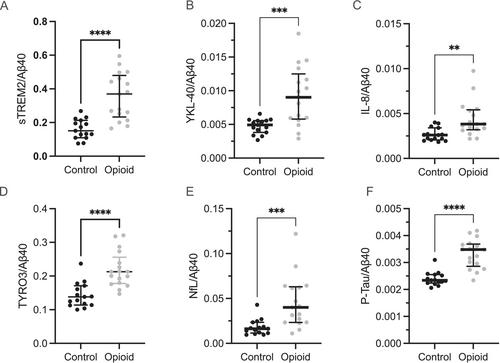
Opioid abuse is a severe global health challenge, leading to rising morbidity, mortality, and increasing societal costs. The aim of this study was to investigate neuroinflammation, neuronal damage and potential changes in the orexin system or beta-amyloid metabolism in the cerebrospinal fluid (CSF) of individuals undergoing opioid substitution therapy (OST). This cross-sectional study investigates CSF biomarkers in individuals undergoing OST, compared to control subjects. Participants receiving OST were recruited from the outpatient clinic at the Department of Psychiatry, Sahlgrenska University Hospital, Gothenburg (Sweden). Each participant provided a complete medical history, including details of drug use over the past 6 months, followed by a lumbar puncture to obtain CSF samples. Molecules associated with neuroinflammation, neuronal and glial damage, beta-amyloid metabolism and orexinergic function were analysed in the participants' CSF, alongside electrolyte levels. Specifically, we analysed levels of sTREM-2, YKL-40, IL-1β, IL-6, IL-8, IL-10, TNF-α, AXL, MER, TYRO3, GAS6, NfL, GFAP, total tau (T-tau), phosphorylated tau (P-tau), neurogranin, Aβ40, Aβ42, the Aβ42/Aβ40 ratio, orexin-A, sPDGFR-β and electrolytes. The study included 15 control subjects and 17 in the opioid substitution group. Patients undergoing opioid substitution therapy exhibited elevated levels of sTREM-2, Aβ42/Aβ40 ratio and NfL in their CSF. Conversely, concentrations of Na+ and Cl− were lower compared to controls. No significant differences were found between groups for other biomarkers, including orexin-A. However, when normalized to Aβ40 levels, YKL-40, IL-8, TYRO3 and P-Tau were also elevated in individuals with opioid dependence. Elevated biomarkers of neuroimmune activation, neuronal damage and beta-amyloid metabolism in opioid dependence suggest CNS inflammation as a contributor to its pathophysiology. Reduced electrolyte levels imply disrupted CSF water regulation, possibly linked to impaired glial function. These findings highlight both neural and non-neural mechanisms in opioid dependence.
期刊介绍:
Addiction Biology is focused on neuroscience contributions and it aims to advance our understanding of the action of drugs of abuse and addictive processes. Papers are accepted in both animal experimentation or clinical research. The content is geared towards behavioral, molecular, genetic, biochemical, neuro-biological and pharmacology aspects of these fields.
Addiction Biology includes peer-reviewed original research reports and reviews.
Addiction Biology is published on behalf of the Society for the Study of Addiction to Alcohol and other Drugs (SSA). Members of the Society for the Study of Addiction receive the Journal as part of their annual membership subscription.
 求助内容:
求助内容: 应助结果提醒方式:


