E M van Bussel, L van Marle, J M Bonsel, D de Vrij, H Weinans, R Sakkers
{"title":"基于超声的统计形状模型对不稳定髋发育不良患者预后的影响。","authors":"E M van Bussel, L van Marle, J M Bonsel, D de Vrij, H Weinans, R Sakkers","doi":"10.1186/s13089-025-00419-3","DOIUrl":null,"url":null,"abstract":"<p><strong>Background: </strong>Current methods to classify developmental dysplasia of the hip (DDH) on ultrasound (US) images, such as the Graf method, provide limited prognostic information. This study aimed to improve the prediction of the clinical course and outcome at age five of decentered hips, diagnosed on the first US made in the first months after birth, by identifying acetabular shape variants on these US images using a statistical shape model (SSM).</p><p><strong>Patients and methods: </strong>US images of the hip were retrieved from a single-center retrospective cohort of patients with DDH Graf type D/III/IV. A SSM was created from the US images made at initial diagnosis.. The association between the identified acetabular shape variants and an unfavorable outcome (residual DDH at age five and open reduction and/or a pelvic osteotomy before age five) was established with multivariable regression models.</p><p><strong>Results: </strong>92 decentered dysplastic hips with full history could be retrieved from the database and were included. At age five, 12 patients (13%) had undergone open reduction, 13 (14%) had a pelvic osteotomy, and 32 (35%) patients showed residual DDH. Four shape variants represented 95% of the variance in acetabular shape. Mode 4 was associated with an unfavorable outcome (odds ratio (OR): 1.80 (95% CI 1.12-2.90). Mode 1 was associated with less risk on open reductions or pelvic osteotomies (OR: 0.56 (95% CI 0.33-0.96).</p><p><strong>Conclusions: </strong>A potential new method of analyzing US images for DDH using SSM established four distinct acetabular shapes on neonatal US images with unstable DDH, of which two were associated with outcomes at five years of age. This tool could serve as a basis for a better prediction of outcome and a more personalized and effective guide for treatment.</p>","PeriodicalId":36911,"journal":{"name":"Ultrasound Journal","volume":"17 1","pages":"26"},"PeriodicalIF":2.9000,"publicationDate":"2025-05-30","publicationTypes":"Journal Article","fieldsOfStudy":null,"isOpenAccess":false,"openAccessPdf":"https://www.ncbi.nlm.nih.gov/pmc/articles/PMC12125406/pdf/","citationCount":"0","resultStr":"{\"title\":\"Ultrasound-based statistical shape modeling for prognosis in unstable hip dysplasia.\",\"authors\":\"E M van Bussel, L van Marle, J M Bonsel, D de Vrij, H Weinans, R Sakkers\",\"doi\":\"10.1186/s13089-025-00419-3\",\"DOIUrl\":null,\"url\":null,\"abstract\":\"<p><strong>Background: </strong>Current methods to classify developmental dysplasia of the hip (DDH) on ultrasound (US) images, such as the Graf method, provide limited prognostic information. This study aimed to improve the prediction of the clinical course and outcome at age five of decentered hips, diagnosed on the first US made in the first months after birth, by identifying acetabular shape variants on these US images using a statistical shape model (SSM).</p><p><strong>Patients and methods: </strong>US images of the hip were retrieved from a single-center retrospective cohort of patients with DDH Graf type D/III/IV. A SSM was created from the US images made at initial diagnosis.. The association between the identified acetabular shape variants and an unfavorable outcome (residual DDH at age five and open reduction and/or a pelvic osteotomy before age five) was established with multivariable regression models.</p><p><strong>Results: </strong>92 decentered dysplastic hips with full history could be retrieved from the database and were included. At age five, 12 patients (13%) had undergone open reduction, 13 (14%) had a pelvic osteotomy, and 32 (35%) patients showed residual DDH. Four shape variants represented 95% of the variance in acetabular shape. Mode 4 was associated with an unfavorable outcome (odds ratio (OR): 1.80 (95% CI 1.12-2.90). Mode 1 was associated with less risk on open reductions or pelvic osteotomies (OR: 0.56 (95% CI 0.33-0.96).</p><p><strong>Conclusions: </strong>A potential new method of analyzing US images for DDH using SSM established four distinct acetabular shapes on neonatal US images with unstable DDH, of which two were associated with outcomes at five years of age. This tool could serve as a basis for a better prediction of outcome and a more personalized and effective guide for treatment.</p>\",\"PeriodicalId\":36911,\"journal\":{\"name\":\"Ultrasound Journal\",\"volume\":\"17 1\",\"pages\":\"26\"},\"PeriodicalIF\":2.9000,\"publicationDate\":\"2025-05-30\",\"publicationTypes\":\"Journal Article\",\"fieldsOfStudy\":null,\"isOpenAccess\":false,\"openAccessPdf\":\"https://www.ncbi.nlm.nih.gov/pmc/articles/PMC12125406/pdf/\",\"citationCount\":\"0\",\"resultStr\":null,\"platform\":\"Semanticscholar\",\"paperid\":null,\"PeriodicalName\":\"Ultrasound Journal\",\"FirstCategoryId\":\"1085\",\"ListUrlMain\":\"https://doi.org/10.1186/s13089-025-00419-3\",\"RegionNum\":0,\"RegionCategory\":null,\"ArticlePicture\":[],\"TitleCN\":null,\"AbstractTextCN\":null,\"PMCID\":null,\"EPubDate\":\"\",\"PubModel\":\"\",\"JCR\":\"Q2\",\"JCRName\":\"Medicine\",\"Score\":null,\"Total\":0}","platform":"Semanticscholar","paperid":null,"PeriodicalName":"Ultrasound Journal","FirstCategoryId":"1085","ListUrlMain":"https://doi.org/10.1186/s13089-025-00419-3","RegionNum":0,"RegionCategory":null,"ArticlePicture":[],"TitleCN":null,"AbstractTextCN":null,"PMCID":null,"EPubDate":"","PubModel":"","JCR":"Q2","JCRName":"Medicine","Score":null,"Total":0}
引用次数: 0
摘要
背景:目前在超声(US)图像上对髋关节发育不良(DDH)进行分类的方法,如Graf方法,提供的预后信息有限。本研究旨在通过使用统计形状模型(SSM)识别这些超声图像上的髋臼形状变异,提高对出生后第一个月进行首次超声诊断的5岁偏心髋的临床病程和结果的预测。患者和方法:从D/III/IV型DDH Graf患者的单中心回顾性队列中检索髋关节超声图像。SSM是根据最初诊断时的美国图像创建的。已确定的髋臼形状变异与不良预后(5岁时残留DDH和5岁前切开复位和/或骨盆截骨)之间的关联通过多变量回归模型建立。结果:从数据库中检索到92例有完整病史的偏心发育不良髋。5岁时,12例(13%)患者行切开复位,13例(14%)行盆腔截骨术,32例(35%)患者出现残留DDH。四种形状变异占髋臼形状变异的95%。模式4与不良结局相关(优势比(OR): 1.80 (95% CI 1.12-2.90)。模式1与切开复位或骨盆截骨的风险较低相关(or: 0.56 (95% CI 0.33-0.96)。结论:使用SSM分析DDH超声图像的一种潜在的新方法在新生儿不稳定DDH超声图像上建立了四种不同的髋臼形状,其中两种与5岁时的预后有关。该工具可以作为更好地预测结果的基础,并为治疗提供更个性化和有效的指导。
Ultrasound-based statistical shape modeling for prognosis in unstable hip dysplasia.
Background: Current methods to classify developmental dysplasia of the hip (DDH) on ultrasound (US) images, such as the Graf method, provide limited prognostic information. This study aimed to improve the prediction of the clinical course and outcome at age five of decentered hips, diagnosed on the first US made in the first months after birth, by identifying acetabular shape variants on these US images using a statistical shape model (SSM).
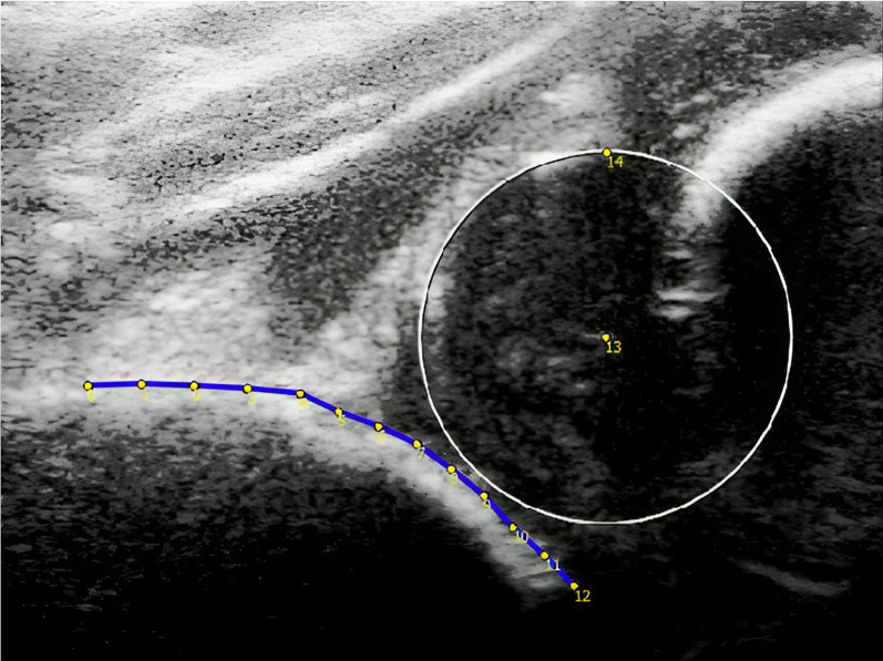
Patients and methods: US images of the hip were retrieved from a single-center retrospective cohort of patients with DDH Graf type D/III/IV. A SSM was created from the US images made at initial diagnosis.. The association between the identified acetabular shape variants and an unfavorable outcome (residual DDH at age five and open reduction and/or a pelvic osteotomy before age five) was established with multivariable regression models.
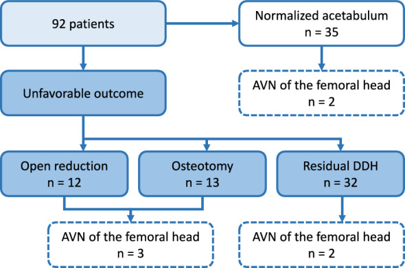
Results: 92 decentered dysplastic hips with full history could be retrieved from the database and were included. At age five, 12 patients (13%) had undergone open reduction, 13 (14%) had a pelvic osteotomy, and 32 (35%) patients showed residual DDH. Four shape variants represented 95% of the variance in acetabular shape. Mode 4 was associated with an unfavorable outcome (odds ratio (OR): 1.80 (95% CI 1.12-2.90). Mode 1 was associated with less risk on open reductions or pelvic osteotomies (OR: 0.56 (95% CI 0.33-0.96).
Conclusions: A potential new method of analyzing US images for DDH using SSM established four distinct acetabular shapes on neonatal US images with unstable DDH, of which two were associated with outcomes at five years of age. This tool could serve as a basis for a better prediction of outcome and a more personalized and effective guide for treatment.


 求助内容:
求助内容: 应助结果提醒方式:


