Ryan M. DeRuiter;Rebecca M. Jones;Hatim Belgharbi;Masashi Sode;Hanjoo R. Lee;Francisco Santibanez;Paul A. Dayton;Gianmarco F. Pinton
{"title":"利用机器人配准复合和非刚性变形校正提高经颅大鼠脑三维超分辨率成像视场。","authors":"Ryan M. DeRuiter;Rebecca M. Jones;Hatim Belgharbi;Masashi Sode;Hanjoo R. Lee;Francisco Santibanez;Paul A. Dayton;Gianmarco F. Pinton","doi":"10.1109/TUFFC.2025.3574916","DOIUrl":null,"url":null,"abstract":"Large field-of-view (FOV) brain imaging with ultrasound has become increasingly achievable with the application of 2-D probes capable of volumetric imaging. However, even in small animals the skull presents a significant barrier and conventional plane-wave transcranial imaging lacks the capability to image in some regions, resulting in incomplete super-resolved vascular reconstructions. Here a high-precision 6 degree-of-freedom robotic approach is used to optimize the transcranial transmission path and to generate composite compounded volumes that improve the field of view and imaging fill fraction. Three-dimensional transcranial simulation quantifies the effect that the skull has on US transmission, and, together with in vivo rat brain results for validation, was used to determine optimal angled transducer orientations for transcranial imaging of ±12°, laterally. Rat brain imaging with an improved FOV was accomplished by a combination of these angles with elevational translations. The 3-D super-resolution results of nine orientations were compounded together using geometric positioning data from the robot arm in combination with a nonrigid deformation correction to account for skull aberration differences. The resulting compounded result was registered against the Waxholm Space rat brain atlas, contextualizing the microvessels. As compared to the zero-angle orientation alone, the compounded result showed improvements in number of vessel-associated voxels for all examined brain regions by at least 350%. Local resolution measurements by a novel 3-D adaptation of a rolling Fourier ring correlation (FRC) approach was used to show consistent resolution measurements between orientation super-resolution results between 10 and <inline-formula> <tex-math>$85~\\mu $ </tex-math></inline-formula>m.","PeriodicalId":13322,"journal":{"name":"IEEE transactions on ultrasonics, ferroelectrics, and frequency control","volume":"72 7","pages":"889-905"},"PeriodicalIF":3.7000,"publicationDate":"2025-03-29","publicationTypes":"Journal Article","fieldsOfStudy":null,"isOpenAccess":false,"openAccessPdf":"","citationCount":"0","resultStr":"{\"title\":\"Improving Imaging Field of View of 3-D Transcranial Rat Brain Super-Resolution With Robotic Registered Compounding and Nonrigid Deformation Correction\",\"authors\":\"Ryan M. DeRuiter;Rebecca M. Jones;Hatim Belgharbi;Masashi Sode;Hanjoo R. Lee;Francisco Santibanez;Paul A. Dayton;Gianmarco F. Pinton\",\"doi\":\"10.1109/TUFFC.2025.3574916\",\"DOIUrl\":null,\"url\":null,\"abstract\":\"Large field-of-view (FOV) brain imaging with ultrasound has become increasingly achievable with the application of 2-D probes capable of volumetric imaging. However, even in small animals the skull presents a significant barrier and conventional plane-wave transcranial imaging lacks the capability to image in some regions, resulting in incomplete super-resolved vascular reconstructions. Here a high-precision 6 degree-of-freedom robotic approach is used to optimize the transcranial transmission path and to generate composite compounded volumes that improve the field of view and imaging fill fraction. Three-dimensional transcranial simulation quantifies the effect that the skull has on US transmission, and, together with in vivo rat brain results for validation, was used to determine optimal angled transducer orientations for transcranial imaging of ±12°, laterally. Rat brain imaging with an improved FOV was accomplished by a combination of these angles with elevational translations. The 3-D super-resolution results of nine orientations were compounded together using geometric positioning data from the robot arm in combination with a nonrigid deformation correction to account for skull aberration differences. The resulting compounded result was registered against the Waxholm Space rat brain atlas, contextualizing the microvessels. As compared to the zero-angle orientation alone, the compounded result showed improvements in number of vessel-associated voxels for all examined brain regions by at least 350%. Local resolution measurements by a novel 3-D adaptation of a rolling Fourier ring correlation (FRC) approach was used to show consistent resolution measurements between orientation super-resolution results between 10 and <inline-formula> <tex-math>$85~\\\\mu $ </tex-math></inline-formula>m.\",\"PeriodicalId\":13322,\"journal\":{\"name\":\"IEEE transactions on ultrasonics, ferroelectrics, and frequency control\",\"volume\":\"72 7\",\"pages\":\"889-905\"},\"PeriodicalIF\":3.7000,\"publicationDate\":\"2025-03-29\",\"publicationTypes\":\"Journal Article\",\"fieldsOfStudy\":null,\"isOpenAccess\":false,\"openAccessPdf\":\"\",\"citationCount\":\"0\",\"resultStr\":null,\"platform\":\"Semanticscholar\",\"paperid\":null,\"PeriodicalName\":\"IEEE transactions on ultrasonics, ferroelectrics, and frequency control\",\"FirstCategoryId\":\"5\",\"ListUrlMain\":\"https://ieeexplore.ieee.org/document/11018119/\",\"RegionNum\":2,\"RegionCategory\":\"工程技术\",\"ArticlePicture\":[],\"TitleCN\":null,\"AbstractTextCN\":null,\"PMCID\":null,\"EPubDate\":\"\",\"PubModel\":\"\",\"JCR\":\"Q1\",\"JCRName\":\"ACOUSTICS\",\"Score\":null,\"Total\":0}","platform":"Semanticscholar","paperid":null,"PeriodicalName":"IEEE transactions on ultrasonics, ferroelectrics, and frequency control","FirstCategoryId":"5","ListUrlMain":"https://ieeexplore.ieee.org/document/11018119/","RegionNum":2,"RegionCategory":"工程技术","ArticlePicture":[],"TitleCN":null,"AbstractTextCN":null,"PMCID":null,"EPubDate":"","PubModel":"","JCR":"Q1","JCRName":"ACOUSTICS","Score":null,"Total":0}
Improving Imaging Field of View of 3-D Transcranial Rat Brain Super-Resolution With Robotic Registered Compounding and Nonrigid Deformation Correction
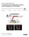
Large field-of-view (FOV) brain imaging with ultrasound has become increasingly achievable with the application of 2-D probes capable of volumetric imaging. However, even in small animals the skull presents a significant barrier and conventional plane-wave transcranial imaging lacks the capability to image in some regions, resulting in incomplete super-resolved vascular reconstructions. Here a high-precision 6 degree-of-freedom robotic approach is used to optimize the transcranial transmission path and to generate composite compounded volumes that improve the field of view and imaging fill fraction. Three-dimensional transcranial simulation quantifies the effect that the skull has on US transmission, and, together with in vivo rat brain results for validation, was used to determine optimal angled transducer orientations for transcranial imaging of ±12°, laterally. Rat brain imaging with an improved FOV was accomplished by a combination of these angles with elevational translations. The 3-D super-resolution results of nine orientations were compounded together using geometric positioning data from the robot arm in combination with a nonrigid deformation correction to account for skull aberration differences. The resulting compounded result was registered against the Waxholm Space rat brain atlas, contextualizing the microvessels. As compared to the zero-angle orientation alone, the compounded result showed improvements in number of vessel-associated voxels for all examined brain regions by at least 350%. Local resolution measurements by a novel 3-D adaptation of a rolling Fourier ring correlation (FRC) approach was used to show consistent resolution measurements between orientation super-resolution results between 10 and $85~\mu $ m.
期刊介绍:
IEEE Transactions on Ultrasonics, Ferroelectrics and Frequency Control includes the theory, technology, materials, and applications relating to: (1) the generation, transmission, and detection of ultrasonic waves and related phenomena; (2) medical ultrasound, including hyperthermia, bioeffects, tissue characterization and imaging; (3) ferroelectric, piezoelectric, and piezomagnetic materials, including crystals, polycrystalline solids, films, polymers, and composites; (4) frequency control, timing and time distribution, including crystal oscillators and other means of classical frequency control, and atomic, molecular and laser frequency control standards. Areas of interest range from fundamental studies to the design and/or applications of devices and systems.