Débora Costa Ruiz, Rocharles Cavalcante Fontenele, Amanda Farias-Gomes, Hugo Gaêta-Araujo, Matheus Lima Oliveira, Deborah Queiroz Freitas, Francisco Haiter-Neto
{"title":"便携式x线仪对近端龋齿病变诊断的影响。","authors":"Débora Costa Ruiz, Rocharles Cavalcante Fontenele, Amanda Farias-Gomes, Hugo Gaêta-Araujo, Matheus Lima Oliveira, Deborah Queiroz Freitas, Francisco Haiter-Neto","doi":"10.1590/1807-3107bor-2025.vol39.028","DOIUrl":null,"url":null,"abstract":"<p><p>This study aimed to evaluate the influence of a portable X-ray device on the diagnosis of proximal caries lesions. For that, radiographs of 40 human teeth with white spots or color changes in enamel and/or dentin were acquired using the Eagle X-ray portable device (Alliage, São Paulo, Brazil) set at 2.5 mA, 60 kVp and an exposure time of 0.5 s (1.25 mAs). Then, new radiographs of the teeth were acquired using the Focus X-ray wall-mounted device (Instrumentarium, Tuusula, Finland) set at 7 mA, 70 kVp, and exposure time of 0.16 s (1.12 mAs). Five oral and maxillofacial radiologists individually assessed the radiographs. Area under the receiver operating characteristic curve (AUC), sensitivity, and specificity were calculated from the responses of the five examiners and compared between the devices tested using Student's t test. Significance level was set at 5% (α = 0.05). The weighted Kappa index evaluated the intra- and inter-examiner agreements for caries lesions diagnosis. The use of a portable X-ray device did not influence on AUC, sensitivity and specificity metrics for the diagnosis of caries lesions (p > 0.05). The intra- and inter-examiner agreements for the caries lesions diagnosis ranged from substantial to almost perfect (0.646-0.859) and moderate to substantial (0.491-0.617), respectively. The diagnostic accuracy for detecting proximal caries lesions is not impaired when using a portable X-ray device.</p>","PeriodicalId":9240,"journal":{"name":"Brazilian oral research","volume":"39 ","pages":"e028"},"PeriodicalIF":1.3000,"publicationDate":"2025-05-23","publicationTypes":"Journal Article","fieldsOfStudy":null,"isOpenAccess":false,"openAccessPdf":"https://www.ncbi.nlm.nih.gov/pmc/articles/PMC12108113/pdf/","citationCount":"0","resultStr":"{\"title\":\"Influence of a portable X-ray device in the diagnosis of proximal caries lesions.\",\"authors\":\"Débora Costa Ruiz, Rocharles Cavalcante Fontenele, Amanda Farias-Gomes, Hugo Gaêta-Araujo, Matheus Lima Oliveira, Deborah Queiroz Freitas, Francisco Haiter-Neto\",\"doi\":\"10.1590/1807-3107bor-2025.vol39.028\",\"DOIUrl\":null,\"url\":null,\"abstract\":\"<p><p>This study aimed to evaluate the influence of a portable X-ray device on the diagnosis of proximal caries lesions. For that, radiographs of 40 human teeth with white spots or color changes in enamel and/or dentin were acquired using the Eagle X-ray portable device (Alliage, São Paulo, Brazil) set at 2.5 mA, 60 kVp and an exposure time of 0.5 s (1.25 mAs). Then, new radiographs of the teeth were acquired using the Focus X-ray wall-mounted device (Instrumentarium, Tuusula, Finland) set at 7 mA, 70 kVp, and exposure time of 0.16 s (1.12 mAs). Five oral and maxillofacial radiologists individually assessed the radiographs. Area under the receiver operating characteristic curve (AUC), sensitivity, and specificity were calculated from the responses of the five examiners and compared between the devices tested using Student's t test. Significance level was set at 5% (α = 0.05). The weighted Kappa index evaluated the intra- and inter-examiner agreements for caries lesions diagnosis. The use of a portable X-ray device did not influence on AUC, sensitivity and specificity metrics for the diagnosis of caries lesions (p > 0.05). The intra- and inter-examiner agreements for the caries lesions diagnosis ranged from substantial to almost perfect (0.646-0.859) and moderate to substantial (0.491-0.617), respectively. The diagnostic accuracy for detecting proximal caries lesions is not impaired when using a portable X-ray device.</p>\",\"PeriodicalId\":9240,\"journal\":{\"name\":\"Brazilian oral research\",\"volume\":\"39 \",\"pages\":\"e028\"},\"PeriodicalIF\":1.3000,\"publicationDate\":\"2025-05-23\",\"publicationTypes\":\"Journal Article\",\"fieldsOfStudy\":null,\"isOpenAccess\":false,\"openAccessPdf\":\"https://www.ncbi.nlm.nih.gov/pmc/articles/PMC12108113/pdf/\",\"citationCount\":\"0\",\"resultStr\":null,\"platform\":\"Semanticscholar\",\"paperid\":null,\"PeriodicalName\":\"Brazilian oral research\",\"FirstCategoryId\":\"3\",\"ListUrlMain\":\"https://doi.org/10.1590/1807-3107bor-2025.vol39.028\",\"RegionNum\":4,\"RegionCategory\":\"医学\",\"ArticlePicture\":[],\"TitleCN\":null,\"AbstractTextCN\":null,\"PMCID\":null,\"EPubDate\":\"2025/1/1 0:00:00\",\"PubModel\":\"eCollection\",\"JCR\":\"Q3\",\"JCRName\":\"DENTISTRY, ORAL SURGERY & MEDICINE\",\"Score\":null,\"Total\":0}","platform":"Semanticscholar","paperid":null,"PeriodicalName":"Brazilian oral research","FirstCategoryId":"3","ListUrlMain":"https://doi.org/10.1590/1807-3107bor-2025.vol39.028","RegionNum":4,"RegionCategory":"医学","ArticlePicture":[],"TitleCN":null,"AbstractTextCN":null,"PMCID":null,"EPubDate":"2025/1/1 0:00:00","PubModel":"eCollection","JCR":"Q3","JCRName":"DENTISTRY, ORAL SURGERY & MEDICINE","Score":null,"Total":0}
引用次数: 0
摘要
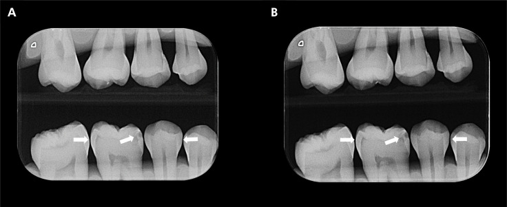
本研究旨在评估便携式x线设备对近端龋齿病变诊断的影响。为此,使用Eagle x射线便携式设备(Alliage, s o Paulo, Brazil),在2.5 mA, 60 kVp和0.5 s (1.25 mA)的曝光时间下,获得了40颗牙釉质和/或牙本质上有白斑或颜色变化的牙齿的x线片。然后,使用Focus x射线壁挂式设备(Instrumentarium, Tuusula, Finland)获得牙齿的新x线片,设置为7 mA, 70 kVp,曝光时间0.16 s (1.12 mA)。5名口腔颌面放射科医生分别评估了x线片。根据五名检查人员的反应计算受试者工作特征曲线下的面积(AUC)、灵敏度和特异性,并使用学生t检验比较测试的设备之间的差异。显著性水平设为5% (α = 0.05)。加权Kappa指数评估内部和内部审查员对龋齿病变诊断的协议。使用便携式x线设备对诊断龋齿的AUC、敏感性和特异性指标没有影响(p < 0.05)。检查人员内部和检查人员之间对龋齿诊断的认同程度从基本到几乎完美(0.646-0.859),中等到基本(0.491-0.617)。当使用便携式x射线设备时,检测近端龋齿病变的诊断准确性不会受到损害。
Influence of a portable X-ray device in the diagnosis of proximal caries lesions.
This study aimed to evaluate the influence of a portable X-ray device on the diagnosis of proximal caries lesions. For that, radiographs of 40 human teeth with white spots or color changes in enamel and/or dentin were acquired using the Eagle X-ray portable device (Alliage, São Paulo, Brazil) set at 2.5 mA, 60 kVp and an exposure time of 0.5 s (1.25 mAs). Then, new radiographs of the teeth were acquired using the Focus X-ray wall-mounted device (Instrumentarium, Tuusula, Finland) set at 7 mA, 70 kVp, and exposure time of 0.16 s (1.12 mAs). Five oral and maxillofacial radiologists individually assessed the radiographs. Area under the receiver operating characteristic curve (AUC), sensitivity, and specificity were calculated from the responses of the five examiners and compared between the devices tested using Student's t test. Significance level was set at 5% (α = 0.05). The weighted Kappa index evaluated the intra- and inter-examiner agreements for caries lesions diagnosis. The use of a portable X-ray device did not influence on AUC, sensitivity and specificity metrics for the diagnosis of caries lesions (p > 0.05). The intra- and inter-examiner agreements for the caries lesions diagnosis ranged from substantial to almost perfect (0.646-0.859) and moderate to substantial (0.491-0.617), respectively. The diagnostic accuracy for detecting proximal caries lesions is not impaired when using a portable X-ray device.
 求助内容:
求助内容: 应助结果提醒方式:


