{"title":"下颌骨消失1例:下颌骨Gorham-Stout病的诊断与治疗。","authors":"Harroun Valdimir T Wong, Johanna Patricia A Cañal","doi":"10.47895/amp.vi0.7516","DOIUrl":null,"url":null,"abstract":"<p><p>Gorham-Stout disease is a rare osteolytic disorder with an unclear pathophysiology. It presents as lesions characterized by the loss of the bony matrix and the proliferation of malformed vasculature. At present, there are no gold-standard diagnostic evaluation protocols and it is diagnosed through a mixture of clinical, histopathologic, and radiographic findings. We report a case of a 19-year-old female with Gorham-Stout disease presenting with an 8-year progressive soft tissue mass in the mandible. Extensive osteolysis of the mandible with clustering of the mandibular dentition is noted on computed tomography (CT) imaging. Her case was discussed in a multidisciplinary conference and her treatment was radiotherapy followed by surgery ± reconstruction. We used a CT-based three-dimensional planning technique to give 40 Gy over 20 treatment sessions to the involved areas. Post treatment, a repeat CT was done at six weeks to reassess for disease progression or stabilization, followed by surgical excision. As of 31 October 2021, no evidence of recurrence is noted 48 months after treatment. Arriving at a definitive diagnosis with Gorham-Stout disease is challenging and a multidisciplinary team approach can help determine the treatment choice with best outcomes.</p>","PeriodicalId":6994,"journal":{"name":"Acta Medica Philippina","volume":"59 5","pages":"75-81"},"PeriodicalIF":0.0000,"publicationDate":"2025-04-30","publicationTypes":"Journal Article","fieldsOfStudy":null,"isOpenAccess":false,"openAccessPdf":"https://www.ncbi.nlm.nih.gov/pmc/articles/PMC12106097/pdf/","citationCount":"0","resultStr":"{\"title\":\"A Case of Vanishing Mandible: Diagnosis and Treatment Considerations for Gorham-Stout Disease of the Mandible.\",\"authors\":\"Harroun Valdimir T Wong, Johanna Patricia A Cañal\",\"doi\":\"10.47895/amp.vi0.7516\",\"DOIUrl\":null,\"url\":null,\"abstract\":\"<p><p>Gorham-Stout disease is a rare osteolytic disorder with an unclear pathophysiology. It presents as lesions characterized by the loss of the bony matrix and the proliferation of malformed vasculature. At present, there are no gold-standard diagnostic evaluation protocols and it is diagnosed through a mixture of clinical, histopathologic, and radiographic findings. We report a case of a 19-year-old female with Gorham-Stout disease presenting with an 8-year progressive soft tissue mass in the mandible. Extensive osteolysis of the mandible with clustering of the mandibular dentition is noted on computed tomography (CT) imaging. Her case was discussed in a multidisciplinary conference and her treatment was radiotherapy followed by surgery ± reconstruction. We used a CT-based three-dimensional planning technique to give 40 Gy over 20 treatment sessions to the involved areas. Post treatment, a repeat CT was done at six weeks to reassess for disease progression or stabilization, followed by surgical excision. As of 31 October 2021, no evidence of recurrence is noted 48 months after treatment. Arriving at a definitive diagnosis with Gorham-Stout disease is challenging and a multidisciplinary team approach can help determine the treatment choice with best outcomes.</p>\",\"PeriodicalId\":6994,\"journal\":{\"name\":\"Acta Medica Philippina\",\"volume\":\"59 5\",\"pages\":\"75-81\"},\"PeriodicalIF\":0.0000,\"publicationDate\":\"2025-04-30\",\"publicationTypes\":\"Journal Article\",\"fieldsOfStudy\":null,\"isOpenAccess\":false,\"openAccessPdf\":\"https://www.ncbi.nlm.nih.gov/pmc/articles/PMC12106097/pdf/\",\"citationCount\":\"0\",\"resultStr\":null,\"platform\":\"Semanticscholar\",\"paperid\":null,\"PeriodicalName\":\"Acta Medica Philippina\",\"FirstCategoryId\":\"1085\",\"ListUrlMain\":\"https://doi.org/10.47895/amp.vi0.7516\",\"RegionNum\":0,\"RegionCategory\":null,\"ArticlePicture\":[],\"TitleCN\":null,\"AbstractTextCN\":null,\"PMCID\":null,\"EPubDate\":\"2025/1/1 0:00:00\",\"PubModel\":\"eCollection\",\"JCR\":\"Q4\",\"JCRName\":\"Medicine\",\"Score\":null,\"Total\":0}","platform":"Semanticscholar","paperid":null,"PeriodicalName":"Acta Medica Philippina","FirstCategoryId":"1085","ListUrlMain":"https://doi.org/10.47895/amp.vi0.7516","RegionNum":0,"RegionCategory":null,"ArticlePicture":[],"TitleCN":null,"AbstractTextCN":null,"PMCID":null,"EPubDate":"2025/1/1 0:00:00","PubModel":"eCollection","JCR":"Q4","JCRName":"Medicine","Score":null,"Total":0}
A Case of Vanishing Mandible: Diagnosis and Treatment Considerations for Gorham-Stout Disease of the Mandible.
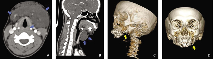
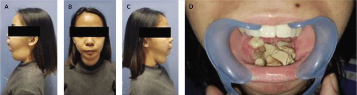
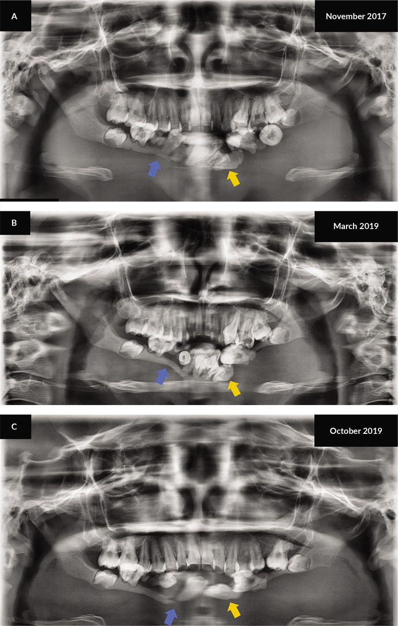
Gorham-Stout disease is a rare osteolytic disorder with an unclear pathophysiology. It presents as lesions characterized by the loss of the bony matrix and the proliferation of malformed vasculature. At present, there are no gold-standard diagnostic evaluation protocols and it is diagnosed through a mixture of clinical, histopathologic, and radiographic findings. We report a case of a 19-year-old female with Gorham-Stout disease presenting with an 8-year progressive soft tissue mass in the mandible. Extensive osteolysis of the mandible with clustering of the mandibular dentition is noted on computed tomography (CT) imaging. Her case was discussed in a multidisciplinary conference and her treatment was radiotherapy followed by surgery ± reconstruction. We used a CT-based three-dimensional planning technique to give 40 Gy over 20 treatment sessions to the involved areas. Post treatment, a repeat CT was done at six weeks to reassess for disease progression or stabilization, followed by surgical excision. As of 31 October 2021, no evidence of recurrence is noted 48 months after treatment. Arriving at a definitive diagnosis with Gorham-Stout disease is challenging and a multidisciplinary team approach can help determine the treatment choice with best outcomes.

 求助内容:
求助内容: 应助结果提醒方式:


