Shang Liu, Zhen Gao, Li-Feng Shi, Han Xiao, Su Li, Meng Li, Zhong-Min Peng
{"title":"基于图像的深度学习模型预测CT≤2 cm肺腺癌淋巴结转移。","authors":"Shang Liu, Zhen Gao, Li-Feng Shi, Han Xiao, Su Li, Meng Li, Zhong-Min Peng","doi":"10.1111/1759-7714.70048","DOIUrl":null,"url":null,"abstract":"<p><strong>Background: </strong>Lymph node metastasis (LNM) poses a considerable threat to survival in lung adenocarcinoma. Currently, minor resection is the recommended surgical approach for small-diameter lung cancer. The accurate preoperative identification of LNM in patients with small-diameter lung cancer is important for improving patient survival and outcomes.</p><p><strong>Methods: </strong>A total of 1740 patients with clinical early-stage lung adenocarcinoma who underwent surgical resection were enrolled in this study. The Lasso model was used to screen clinical and imaging features, and multivariate logistic regression analysis was used to analyze the relevant diagnostic factors to establish a diagnostic model for predicting LNM. Receiver operating characteristic (ROC) curve analysis, decision curve analysis (DCA) and calibration curve analysis were used to verify the clinical efficacy of the model, which was further validated with an internal validation set.</p><p><strong>Results: </strong>The proportion of solid components (PSC), sphericity, nodule margin, entropy, and edge blur were identified as diagnostic factors that were strongly correlated with LNM in lung adenocarcinoma patients. The area under the ROC curve (AUC) in the internal training set was 0.91. Decision curve analysis revealed that the model could achieve greater benefits for patients. The calibration curve was used to further verify the applicability of the prediction model.</p><p><strong>Conclusions: </strong>Patients with early-stage lung adenocarcinoma with LNM can be identified by typical imaging features. The diagnostic model can help to optimize surgical planning among thoracic surgeons.</p>","PeriodicalId":23338,"journal":{"name":"Thoracic Cancer","volume":"16 10","pages":"e70048"},"PeriodicalIF":2.3000,"publicationDate":"2025-05-01","publicationTypes":"Journal Article","fieldsOfStudy":null,"isOpenAccess":false,"openAccessPdf":"https://www.ncbi.nlm.nih.gov/pmc/articles/PMC12116230/pdf/","citationCount":"0","resultStr":"{\"title\":\"Image-Based Deep Learning Model for Predicting Lymph Node Metastasis in Lung Adenocarcinoma With CT ≤ 2 cm.\",\"authors\":\"Shang Liu, Zhen Gao, Li-Feng Shi, Han Xiao, Su Li, Meng Li, Zhong-Min Peng\",\"doi\":\"10.1111/1759-7714.70048\",\"DOIUrl\":null,\"url\":null,\"abstract\":\"<p><strong>Background: </strong>Lymph node metastasis (LNM) poses a considerable threat to survival in lung adenocarcinoma. Currently, minor resection is the recommended surgical approach for small-diameter lung cancer. The accurate preoperative identification of LNM in patients with small-diameter lung cancer is important for improving patient survival and outcomes.</p><p><strong>Methods: </strong>A total of 1740 patients with clinical early-stage lung adenocarcinoma who underwent surgical resection were enrolled in this study. The Lasso model was used to screen clinical and imaging features, and multivariate logistic regression analysis was used to analyze the relevant diagnostic factors to establish a diagnostic model for predicting LNM. Receiver operating characteristic (ROC) curve analysis, decision curve analysis (DCA) and calibration curve analysis were used to verify the clinical efficacy of the model, which was further validated with an internal validation set.</p><p><strong>Results: </strong>The proportion of solid components (PSC), sphericity, nodule margin, entropy, and edge blur were identified as diagnostic factors that were strongly correlated with LNM in lung adenocarcinoma patients. The area under the ROC curve (AUC) in the internal training set was 0.91. Decision curve analysis revealed that the model could achieve greater benefits for patients. The calibration curve was used to further verify the applicability of the prediction model.</p><p><strong>Conclusions: </strong>Patients with early-stage lung adenocarcinoma with LNM can be identified by typical imaging features. The diagnostic model can help to optimize surgical planning among thoracic surgeons.</p>\",\"PeriodicalId\":23338,\"journal\":{\"name\":\"Thoracic Cancer\",\"volume\":\"16 10\",\"pages\":\"e70048\"},\"PeriodicalIF\":2.3000,\"publicationDate\":\"2025-05-01\",\"publicationTypes\":\"Journal Article\",\"fieldsOfStudy\":null,\"isOpenAccess\":false,\"openAccessPdf\":\"https://www.ncbi.nlm.nih.gov/pmc/articles/PMC12116230/pdf/\",\"citationCount\":\"0\",\"resultStr\":null,\"platform\":\"Semanticscholar\",\"paperid\":null,\"PeriodicalName\":\"Thoracic Cancer\",\"FirstCategoryId\":\"3\",\"ListUrlMain\":\"https://doi.org/10.1111/1759-7714.70048\",\"RegionNum\":3,\"RegionCategory\":\"医学\",\"ArticlePicture\":[],\"TitleCN\":null,\"AbstractTextCN\":null,\"PMCID\":null,\"EPubDate\":\"\",\"PubModel\":\"\",\"JCR\":\"Q3\",\"JCRName\":\"ONCOLOGY\",\"Score\":null,\"Total\":0}","platform":"Semanticscholar","paperid":null,"PeriodicalName":"Thoracic Cancer","FirstCategoryId":"3","ListUrlMain":"https://doi.org/10.1111/1759-7714.70048","RegionNum":3,"RegionCategory":"医学","ArticlePicture":[],"TitleCN":null,"AbstractTextCN":null,"PMCID":null,"EPubDate":"","PubModel":"","JCR":"Q3","JCRName":"ONCOLOGY","Score":null,"Total":0}
Image-Based Deep Learning Model for Predicting Lymph Node Metastasis in Lung Adenocarcinoma With CT ≤ 2 cm.
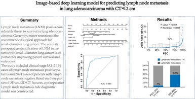
Background: Lymph node metastasis (LNM) poses a considerable threat to survival in lung adenocarcinoma. Currently, minor resection is the recommended surgical approach for small-diameter lung cancer. The accurate preoperative identification of LNM in patients with small-diameter lung cancer is important for improving patient survival and outcomes.
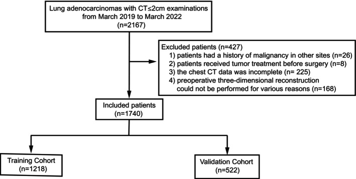
Methods: A total of 1740 patients with clinical early-stage lung adenocarcinoma who underwent surgical resection were enrolled in this study. The Lasso model was used to screen clinical and imaging features, and multivariate logistic regression analysis was used to analyze the relevant diagnostic factors to establish a diagnostic model for predicting LNM. Receiver operating characteristic (ROC) curve analysis, decision curve analysis (DCA) and calibration curve analysis were used to verify the clinical efficacy of the model, which was further validated with an internal validation set.
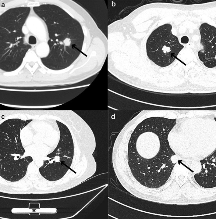
Results: The proportion of solid components (PSC), sphericity, nodule margin, entropy, and edge blur were identified as diagnostic factors that were strongly correlated with LNM in lung adenocarcinoma patients. The area under the ROC curve (AUC) in the internal training set was 0.91. Decision curve analysis revealed that the model could achieve greater benefits for patients. The calibration curve was used to further verify the applicability of the prediction model.
Conclusions: Patients with early-stage lung adenocarcinoma with LNM can be identified by typical imaging features. The diagnostic model can help to optimize surgical planning among thoracic surgeons.
期刊介绍:
Thoracic Cancer aims to facilitate international collaboration and exchange of comprehensive and cutting-edge information on basic, translational, and applied clinical research in lung cancer, esophageal cancer, mediastinal cancer, breast cancer and other thoracic malignancies. Prevention, treatment and research relevant to Asia-Pacific is a focus area, but submissions from all regions are welcomed. The editors encourage contributions relevant to prevention, general thoracic surgery, medical oncology, radiology, radiation medicine, pathology, basic cancer research, as well as epidemiological and translational studies in thoracic cancer. Thoracic Cancer is the official publication of the Chinese Society of Lung Cancer, International Chinese Society of Thoracic Surgery and is endorsed by the Korean Association for the Study of Lung Cancer and the Hong Kong Cancer Therapy Society.
The Journal publishes a range of article types including: Editorials, Invited Reviews, Mini Reviews, Original Articles, Clinical Guidelines, Technological Notes, Imaging in thoracic cancer, Meeting Reports, Case Reports, Letters to the Editor, Commentaries, and Brief Reports.
 求助内容:
求助内容: 应助结果提醒方式:


