Lei Chen, Bowen Qiu, Xinjia Du, Jiahua Liu, Zhongxiao Liu, Wensu Chen, Wenliang Che, Yuan Lu
{"title":"心脏MRI评估的细胞外体积与st段抬高型心肌梗死患者新发心房颤动的关系","authors":"Lei Chen, Bowen Qiu, Xinjia Du, Jiahua Liu, Zhongxiao Liu, Wensu Chen, Wenliang Che, Yuan Lu","doi":"10.3348/kjr.2025.0070","DOIUrl":null,"url":null,"abstract":"<p><strong>Objective: </strong>Although left ventricular (LV) fibrosis has been strongly linked to atrial fibrillation (AF), its relationship with new-onset AF (NOAF) following ST-segment elevation myocardial infarction (STEMI) remains unclear. This study aimed to investigate the association between different extracellular volume (ECV) measurements in the LV and NOAF during acute-phase STEMI.</p><p><strong>Materials and methods: </strong>This retrospective study included 517 patients diagnosed with acute STEMI (440 males, 77 females; mean age, 57.2 ± 12.4 years). All patients underwent cardiac magnetic resonance (CMR) imaging with T1 mapping sequences during hospitalization. Blood samples were collected within 24 hours of the CMR examination. ECV was assessed in three regions of the left ventricle: the non-myocardial infarction region (NMI-ECV), the myocardial infarction region (MI-ECV), and the entire myocardium (integral ECV). Multi-variable logistic regression was used to evaluate the associations between these ECV parameters and NOAF. Receiver operating characteristic (ROC) analysis was performed to assess the predictive value of ECV measurements, both alone and in combination with two conventional risk factors-N-terminal pro-B-type natriuretic peptide and infarct-related artery (right coronary artery).</p><p><strong>Results: </strong>During hospitalization, 40 (7.7%) patients developed NOAF. After adjusting for confounding factors, including left atrial strain, MI-ECV, NMI-ECV, and integral ECV were independently associated with NOAF. The area under the ROC curve for predicting NOAF was 0.702 (95% confidence interval: 0.615-0.789), 0.625 (0.531-0.719), and 0.712 (0.627-0.798) for MI-ECV, NMI-ECV, and integral ECV, respectively. The addition of integral ECV and MI-ECV to conventional factors significantly improved the predictive performance for NOAF.</p><p><strong>Conclusion: </strong>ECV measured using CMR was independently and significantly associated with NOAF occurrence in acute-phase STEMI. Incorporating ECV into risk assessment models could significantly improve NOAF prediction.</p>","PeriodicalId":17881,"journal":{"name":"Korean Journal of Radiology","volume":"26 6","pages":"546-556"},"PeriodicalIF":5.3000,"publicationDate":"2025-06-01","publicationTypes":"Journal Article","fieldsOfStudy":null,"isOpenAccess":false,"openAccessPdf":"https://www.ncbi.nlm.nih.gov/pmc/articles/PMC12123073/pdf/","citationCount":"0","resultStr":"{\"title\":\"Association Between Extracellular Volume Assessed by Cardiac MRI and New-Onset Atrial Fibrillation in Patients With ST-Segment Elevation Myocardial Infarction.\",\"authors\":\"Lei Chen, Bowen Qiu, Xinjia Du, Jiahua Liu, Zhongxiao Liu, Wensu Chen, Wenliang Che, Yuan Lu\",\"doi\":\"10.3348/kjr.2025.0070\",\"DOIUrl\":null,\"url\":null,\"abstract\":\"<p><strong>Objective: </strong>Although left ventricular (LV) fibrosis has been strongly linked to atrial fibrillation (AF), its relationship with new-onset AF (NOAF) following ST-segment elevation myocardial infarction (STEMI) remains unclear. This study aimed to investigate the association between different extracellular volume (ECV) measurements in the LV and NOAF during acute-phase STEMI.</p><p><strong>Materials and methods: </strong>This retrospective study included 517 patients diagnosed with acute STEMI (440 males, 77 females; mean age, 57.2 ± 12.4 years). All patients underwent cardiac magnetic resonance (CMR) imaging with T1 mapping sequences during hospitalization. Blood samples were collected within 24 hours of the CMR examination. ECV was assessed in three regions of the left ventricle: the non-myocardial infarction region (NMI-ECV), the myocardial infarction region (MI-ECV), and the entire myocardium (integral ECV). Multi-variable logistic regression was used to evaluate the associations between these ECV parameters and NOAF. Receiver operating characteristic (ROC) analysis was performed to assess the predictive value of ECV measurements, both alone and in combination with two conventional risk factors-N-terminal pro-B-type natriuretic peptide and infarct-related artery (right coronary artery).</p><p><strong>Results: </strong>During hospitalization, 40 (7.7%) patients developed NOAF. After adjusting for confounding factors, including left atrial strain, MI-ECV, NMI-ECV, and integral ECV were independently associated with NOAF. The area under the ROC curve for predicting NOAF was 0.702 (95% confidence interval: 0.615-0.789), 0.625 (0.531-0.719), and 0.712 (0.627-0.798) for MI-ECV, NMI-ECV, and integral ECV, respectively. The addition of integral ECV and MI-ECV to conventional factors significantly improved the predictive performance for NOAF.</p><p><strong>Conclusion: </strong>ECV measured using CMR was independently and significantly associated with NOAF occurrence in acute-phase STEMI. Incorporating ECV into risk assessment models could significantly improve NOAF prediction.</p>\",\"PeriodicalId\":17881,\"journal\":{\"name\":\"Korean Journal of Radiology\",\"volume\":\"26 6\",\"pages\":\"546-556\"},\"PeriodicalIF\":5.3000,\"publicationDate\":\"2025-06-01\",\"publicationTypes\":\"Journal Article\",\"fieldsOfStudy\":null,\"isOpenAccess\":false,\"openAccessPdf\":\"https://www.ncbi.nlm.nih.gov/pmc/articles/PMC12123073/pdf/\",\"citationCount\":\"0\",\"resultStr\":null,\"platform\":\"Semanticscholar\",\"paperid\":null,\"PeriodicalName\":\"Korean Journal of Radiology\",\"FirstCategoryId\":\"3\",\"ListUrlMain\":\"https://doi.org/10.3348/kjr.2025.0070\",\"RegionNum\":2,\"RegionCategory\":\"医学\",\"ArticlePicture\":[],\"TitleCN\":null,\"AbstractTextCN\":null,\"PMCID\":null,\"EPubDate\":\"\",\"PubModel\":\"\",\"JCR\":\"Q1\",\"JCRName\":\"RADIOLOGY, NUCLEAR MEDICINE & MEDICAL IMAGING\",\"Score\":null,\"Total\":0}","platform":"Semanticscholar","paperid":null,"PeriodicalName":"Korean Journal of Radiology","FirstCategoryId":"3","ListUrlMain":"https://doi.org/10.3348/kjr.2025.0070","RegionNum":2,"RegionCategory":"医学","ArticlePicture":[],"TitleCN":null,"AbstractTextCN":null,"PMCID":null,"EPubDate":"","PubModel":"","JCR":"Q1","JCRName":"RADIOLOGY, NUCLEAR MEDICINE & MEDICAL IMAGING","Score":null,"Total":0}
Association Between Extracellular Volume Assessed by Cardiac MRI and New-Onset Atrial Fibrillation in Patients With ST-Segment Elevation Myocardial Infarction.
Objective: Although left ventricular (LV) fibrosis has been strongly linked to atrial fibrillation (AF), its relationship with new-onset AF (NOAF) following ST-segment elevation myocardial infarction (STEMI) remains unclear. This study aimed to investigate the association between different extracellular volume (ECV) measurements in the LV and NOAF during acute-phase STEMI.
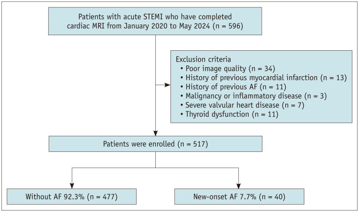
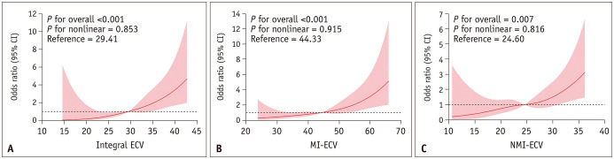
Materials and methods: This retrospective study included 517 patients diagnosed with acute STEMI (440 males, 77 females; mean age, 57.2 ± 12.4 years). All patients underwent cardiac magnetic resonance (CMR) imaging with T1 mapping sequences during hospitalization. Blood samples were collected within 24 hours of the CMR examination. ECV was assessed in three regions of the left ventricle: the non-myocardial infarction region (NMI-ECV), the myocardial infarction region (MI-ECV), and the entire myocardium (integral ECV). Multi-variable logistic regression was used to evaluate the associations between these ECV parameters and NOAF. Receiver operating characteristic (ROC) analysis was performed to assess the predictive value of ECV measurements, both alone and in combination with two conventional risk factors-N-terminal pro-B-type natriuretic peptide and infarct-related artery (right coronary artery).
Results: During hospitalization, 40 (7.7%) patients developed NOAF. After adjusting for confounding factors, including left atrial strain, MI-ECV, NMI-ECV, and integral ECV were independently associated with NOAF. The area under the ROC curve for predicting NOAF was 0.702 (95% confidence interval: 0.615-0.789), 0.625 (0.531-0.719), and 0.712 (0.627-0.798) for MI-ECV, NMI-ECV, and integral ECV, respectively. The addition of integral ECV and MI-ECV to conventional factors significantly improved the predictive performance for NOAF.
Conclusion: ECV measured using CMR was independently and significantly associated with NOAF occurrence in acute-phase STEMI. Incorporating ECV into risk assessment models could significantly improve NOAF prediction.
期刊介绍:
The inaugural issue of the Korean J Radiol came out in March 2000. Our journal aims to produce and propagate knowledge on radiologic imaging and related sciences.
A unique feature of the articles published in the Journal will be their reflection of global trends in radiology combined with an East-Asian perspective. Geographic differences in disease prevalence will be reflected in the contents of papers, and this will serve to enrich our body of knowledge.
World''s outstanding radiologists from many countries are serving as editorial board of our journal.


 求助内容:
求助内容: 应助结果提醒方式:


