Dina Soliman, Firyal Ibrahim, Hasan Rizvi, Mahir Petkar, Zsolt Lengyel, Aliya Habib Sange, Awni Alshurafa, Ruba Yasin
{"title":"BRAF v660e突变双表型Erdheim-Chester肿瘤/ rossai - dorfman病(混合性组织细胞肿瘤)具有不典型组织学特征和暴发性噬血症,广泛累及骨髓","authors":"Dina Soliman, Firyal Ibrahim, Hasan Rizvi, Mahir Petkar, Zsolt Lengyel, Aliya Habib Sange, Awni Alshurafa, Ruba Yasin","doi":"10.1159/000545775","DOIUrl":null,"url":null,"abstract":"<p><strong>Background: </strong>Erdheim-Chester disease (ECD) is a recently recognized clonal hematopoietic neoplasm characterized by activating alterations in the MAPK pathway. It involves multi-organ accumulation of abnormal histiocytes, leading to nonspecific clinical manifestations due to inflammation and fibrosis caused by histiocytic infiltration.</p><p><strong>Case presentation: </strong>We present a 45-year-old male with nonspecific clinical symptoms and progressive skin and abdominal lesions. Multiple tissue biopsies revealed fibrohistiocytic infiltration but provided an inconclusive diagnosis. Imaging studies showed extensive fibrosis in the perinephric regions on CT and sclerotic foci in long and pelvic bones on PET/CT. Bone marrow biopsy revealed abnormal histiocytes with multinucleated giant forms, prominent emperipolesis, active hemophagocytosis, and condensed hemosiderin deposition. Immunohistochemistry showed positive histiocytes for CD68, CD163, and partially for S-100. Molecular analysis confirmed the <i>BRAFV660E</i> mutation, establishing a diagnosis of ECD with atypical histologic features and findings overlapping with Rosai-Dorfman (mixed histiocytosis). The diagnosis was challenging due to extensive fibrosis, the lack of typical histopathologic features of ECD, in addition to concurrent involvement by Rosai-Dorfman cells (mixed histiocytosis), activated macrophages, and dense hemosiderin deposition - a morphologic characteristic not previously described in ECD. Unfortunately, the diagnosis was delayed by 6 years, which tragically led to a fatal outcome.</p><p><strong>Conclusion: </strong>The case highlights the need to recognize that ECD diagnosis requires integrating histopathology with clinical and radiographic findings. It emphasizes the importance of awareness of mixed histiocytosis features and the role of detecting BRAF mutations, even through immunohistochemistry, in suspected histiocytic neoplasms. Extensive bone marrow involvement by ECD is rarely described. To our knowledge, there are no prior reports of bi-phenotypic (concurrent) ECD/RDD mixed histiocytosis affecting the bone marrow.</p>","PeriodicalId":9625,"journal":{"name":"Case Reports in Oncology","volume":"18 1","pages":"602-612"},"PeriodicalIF":0.7000,"publicationDate":"2025-05-26","publicationTypes":"Journal Article","fieldsOfStudy":null,"isOpenAccess":false,"openAccessPdf":"https://www.ncbi.nlm.nih.gov/pmc/articles/PMC12105831/pdf/","citationCount":"0","resultStr":"{\"title\":\"Extensive Bone Marrow Involvement by <i>BRAF</i> <sup>V660E</sup>-Mutated Bi-Phenotypic Erdheim-Chester Neoplasm/Rosai-Dorfman Disease (Mixed Histiocytic Neoplasm) with Atypical Histological Features and Fulminant Hemophagocytosis.\",\"authors\":\"Dina Soliman, Firyal Ibrahim, Hasan Rizvi, Mahir Petkar, Zsolt Lengyel, Aliya Habib Sange, Awni Alshurafa, Ruba Yasin\",\"doi\":\"10.1159/000545775\",\"DOIUrl\":null,\"url\":null,\"abstract\":\"<p><strong>Background: </strong>Erdheim-Chester disease (ECD) is a recently recognized clonal hematopoietic neoplasm characterized by activating alterations in the MAPK pathway. It involves multi-organ accumulation of abnormal histiocytes, leading to nonspecific clinical manifestations due to inflammation and fibrosis caused by histiocytic infiltration.</p><p><strong>Case presentation: </strong>We present a 45-year-old male with nonspecific clinical symptoms and progressive skin and abdominal lesions. Multiple tissue biopsies revealed fibrohistiocytic infiltration but provided an inconclusive diagnosis. Imaging studies showed extensive fibrosis in the perinephric regions on CT and sclerotic foci in long and pelvic bones on PET/CT. Bone marrow biopsy revealed abnormal histiocytes with multinucleated giant forms, prominent emperipolesis, active hemophagocytosis, and condensed hemosiderin deposition. Immunohistochemistry showed positive histiocytes for CD68, CD163, and partially for S-100. Molecular analysis confirmed the <i>BRAFV660E</i> mutation, establishing a diagnosis of ECD with atypical histologic features and findings overlapping with Rosai-Dorfman (mixed histiocytosis). The diagnosis was challenging due to extensive fibrosis, the lack of typical histopathologic features of ECD, in addition to concurrent involvement by Rosai-Dorfman cells (mixed histiocytosis), activated macrophages, and dense hemosiderin deposition - a morphologic characteristic not previously described in ECD. Unfortunately, the diagnosis was delayed by 6 years, which tragically led to a fatal outcome.</p><p><strong>Conclusion: </strong>The case highlights the need to recognize that ECD diagnosis requires integrating histopathology with clinical and radiographic findings. It emphasizes the importance of awareness of mixed histiocytosis features and the role of detecting BRAF mutations, even through immunohistochemistry, in suspected histiocytic neoplasms. Extensive bone marrow involvement by ECD is rarely described. To our knowledge, there are no prior reports of bi-phenotypic (concurrent) ECD/RDD mixed histiocytosis affecting the bone marrow.</p>\",\"PeriodicalId\":9625,\"journal\":{\"name\":\"Case Reports in Oncology\",\"volume\":\"18 1\",\"pages\":\"602-612\"},\"PeriodicalIF\":0.7000,\"publicationDate\":\"2025-05-26\",\"publicationTypes\":\"Journal Article\",\"fieldsOfStudy\":null,\"isOpenAccess\":false,\"openAccessPdf\":\"https://www.ncbi.nlm.nih.gov/pmc/articles/PMC12105831/pdf/\",\"citationCount\":\"0\",\"resultStr\":null,\"platform\":\"Semanticscholar\",\"paperid\":null,\"PeriodicalName\":\"Case Reports in Oncology\",\"FirstCategoryId\":\"1085\",\"ListUrlMain\":\"https://doi.org/10.1159/000545775\",\"RegionNum\":0,\"RegionCategory\":null,\"ArticlePicture\":[],\"TitleCN\":null,\"AbstractTextCN\":null,\"PMCID\":null,\"EPubDate\":\"2025/1/1 0:00:00\",\"PubModel\":\"eCollection\",\"JCR\":\"Q4\",\"JCRName\":\"ONCOLOGY\",\"Score\":null,\"Total\":0}","platform":"Semanticscholar","paperid":null,"PeriodicalName":"Case Reports in Oncology","FirstCategoryId":"1085","ListUrlMain":"https://doi.org/10.1159/000545775","RegionNum":0,"RegionCategory":null,"ArticlePicture":[],"TitleCN":null,"AbstractTextCN":null,"PMCID":null,"EPubDate":"2025/1/1 0:00:00","PubModel":"eCollection","JCR":"Q4","JCRName":"ONCOLOGY","Score":null,"Total":0}
Extensive Bone Marrow Involvement by BRAFV660E-Mutated Bi-Phenotypic Erdheim-Chester Neoplasm/Rosai-Dorfman Disease (Mixed Histiocytic Neoplasm) with Atypical Histological Features and Fulminant Hemophagocytosis.
Background: Erdheim-Chester disease (ECD) is a recently recognized clonal hematopoietic neoplasm characterized by activating alterations in the MAPK pathway. It involves multi-organ accumulation of abnormal histiocytes, leading to nonspecific clinical manifestations due to inflammation and fibrosis caused by histiocytic infiltration.
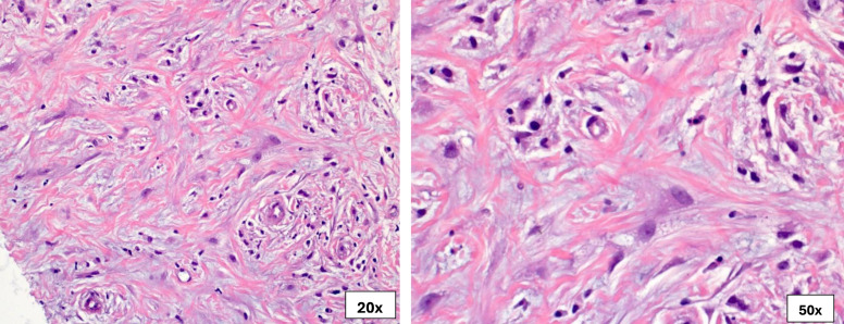
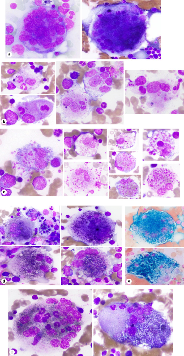
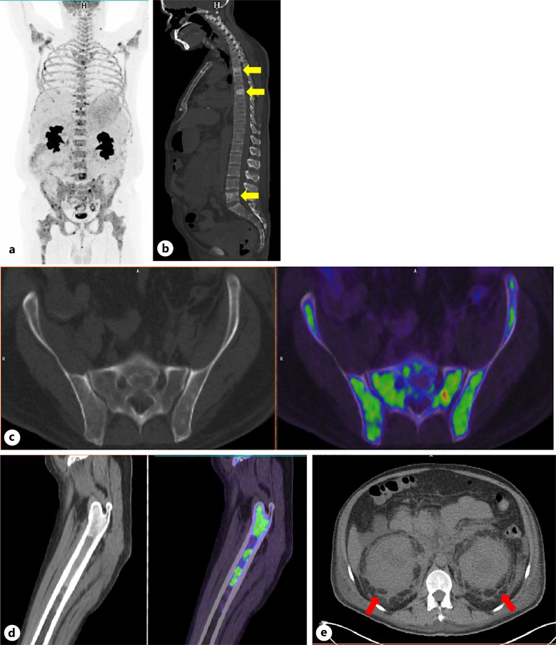
Case presentation: We present a 45-year-old male with nonspecific clinical symptoms and progressive skin and abdominal lesions. Multiple tissue biopsies revealed fibrohistiocytic infiltration but provided an inconclusive diagnosis. Imaging studies showed extensive fibrosis in the perinephric regions on CT and sclerotic foci in long and pelvic bones on PET/CT. Bone marrow biopsy revealed abnormal histiocytes with multinucleated giant forms, prominent emperipolesis, active hemophagocytosis, and condensed hemosiderin deposition. Immunohistochemistry showed positive histiocytes for CD68, CD163, and partially for S-100. Molecular analysis confirmed the BRAFV660E mutation, establishing a diagnosis of ECD with atypical histologic features and findings overlapping with Rosai-Dorfman (mixed histiocytosis). The diagnosis was challenging due to extensive fibrosis, the lack of typical histopathologic features of ECD, in addition to concurrent involvement by Rosai-Dorfman cells (mixed histiocytosis), activated macrophages, and dense hemosiderin deposition - a morphologic characteristic not previously described in ECD. Unfortunately, the diagnosis was delayed by 6 years, which tragically led to a fatal outcome.
Conclusion: The case highlights the need to recognize that ECD diagnosis requires integrating histopathology with clinical and radiographic findings. It emphasizes the importance of awareness of mixed histiocytosis features and the role of detecting BRAF mutations, even through immunohistochemistry, in suspected histiocytic neoplasms. Extensive bone marrow involvement by ECD is rarely described. To our knowledge, there are no prior reports of bi-phenotypic (concurrent) ECD/RDD mixed histiocytosis affecting the bone marrow.

 求助内容:
求助内容: 应助结果提醒方式:


