{"title":"综合后向散射血管内超声评价冠状动脉造影黄色分级与脂质斑块的相关性。","authors":"Atsushi Tanita, Shinichiro Sunamura, Tsuyoshi Ogata, Kazuki Noda, Toru Takii, Yoshio Nitta, Seijiro Yoshida, Shigeto Namiuchi","doi":"10.1007/s12928-025-01133-6","DOIUrl":null,"url":null,"abstract":"<p><p>Coronary angioscopy (CAS) enables direct qualitative assessment of the coronary artery lumen, while integrated backscatter intravascular ultrasound (IB-IVUS) provides a quantitative evaluation of coronary plaque tissue characteristics. Despite the utility of both techniques in assessing coronary plaque status, data on the correlation between their findings remain limited. To investigate the association between CAS-derived findings and results obtained through IB-IVUS. This retrospective analysis included 36 patients who underwent both CAS and IB-IVUS during percutaneous coronary intervention (PCI) at our institution. CAS and IB-IVUS were performed on the same coronary artery treated during PCI. Plaques were categorized into four groups based on their yellow color grade using CAS. For the IB-IVUS analysis, measurements were performed at the minimum lumen diameter site of the culprit lesion. A significant correlation was observed between plaque yellowishness and plaque characteristics on IB-IVUS. Higher plaque yellowishness was associated with an increased percentage of all lipid pool (P < 0.01), a greater proportion of attenuated plaque (P < 0.01), and a larger estimated lipid plaque volume (P < 0.01). Additionally, plaques with higher yellowishness grades had significantly thinner fibrous caps (P < 0.01). The findings suggest that higher plaque yellowishness observed via CAS correlates with a larger lipid plaque volume and thinner fibrous caps, as assessed through IB-IVUS.</p>","PeriodicalId":9439,"journal":{"name":"Cardiovascular Intervention and Therapeutics","volume":" ","pages":"778-787"},"PeriodicalIF":5.8000,"publicationDate":"2025-10-01","publicationTypes":"Journal Article","fieldsOfStudy":null,"isOpenAccess":false,"openAccessPdf":"https://www.ncbi.nlm.nih.gov/pmc/articles/PMC12432081/pdf/","citationCount":"0","resultStr":"{\"title\":\"Correlation between coronary angioscopy yellow grade and lipid plaque assessment by integrated backscatter intravascular ultrasound.\",\"authors\":\"Atsushi Tanita, Shinichiro Sunamura, Tsuyoshi Ogata, Kazuki Noda, Toru Takii, Yoshio Nitta, Seijiro Yoshida, Shigeto Namiuchi\",\"doi\":\"10.1007/s12928-025-01133-6\",\"DOIUrl\":null,\"url\":null,\"abstract\":\"<p><p>Coronary angioscopy (CAS) enables direct qualitative assessment of the coronary artery lumen, while integrated backscatter intravascular ultrasound (IB-IVUS) provides a quantitative evaluation of coronary plaque tissue characteristics. Despite the utility of both techniques in assessing coronary plaque status, data on the correlation between their findings remain limited. To investigate the association between CAS-derived findings and results obtained through IB-IVUS. This retrospective analysis included 36 patients who underwent both CAS and IB-IVUS during percutaneous coronary intervention (PCI) at our institution. CAS and IB-IVUS were performed on the same coronary artery treated during PCI. Plaques were categorized into four groups based on their yellow color grade using CAS. For the IB-IVUS analysis, measurements were performed at the minimum lumen diameter site of the culprit lesion. A significant correlation was observed between plaque yellowishness and plaque characteristics on IB-IVUS. Higher plaque yellowishness was associated with an increased percentage of all lipid pool (P < 0.01), a greater proportion of attenuated plaque (P < 0.01), and a larger estimated lipid plaque volume (P < 0.01). Additionally, plaques with higher yellowishness grades had significantly thinner fibrous caps (P < 0.01). The findings suggest that higher plaque yellowishness observed via CAS correlates with a larger lipid plaque volume and thinner fibrous caps, as assessed through IB-IVUS.</p>\",\"PeriodicalId\":9439,\"journal\":{\"name\":\"Cardiovascular Intervention and Therapeutics\",\"volume\":\" \",\"pages\":\"778-787\"},\"PeriodicalIF\":5.8000,\"publicationDate\":\"2025-10-01\",\"publicationTypes\":\"Journal Article\",\"fieldsOfStudy\":null,\"isOpenAccess\":false,\"openAccessPdf\":\"https://www.ncbi.nlm.nih.gov/pmc/articles/PMC12432081/pdf/\",\"citationCount\":\"0\",\"resultStr\":null,\"platform\":\"Semanticscholar\",\"paperid\":null,\"PeriodicalName\":\"Cardiovascular Intervention and Therapeutics\",\"FirstCategoryId\":\"1085\",\"ListUrlMain\":\"https://doi.org/10.1007/s12928-025-01133-6\",\"RegionNum\":0,\"RegionCategory\":null,\"ArticlePicture\":[],\"TitleCN\":null,\"AbstractTextCN\":null,\"PMCID\":null,\"EPubDate\":\"2025/5/27 0:00:00\",\"PubModel\":\"Epub\",\"JCR\":\"Q2\",\"JCRName\":\"CARDIAC & CARDIOVASCULAR SYSTEMS\",\"Score\":null,\"Total\":0}","platform":"Semanticscholar","paperid":null,"PeriodicalName":"Cardiovascular Intervention and Therapeutics","FirstCategoryId":"1085","ListUrlMain":"https://doi.org/10.1007/s12928-025-01133-6","RegionNum":0,"RegionCategory":null,"ArticlePicture":[],"TitleCN":null,"AbstractTextCN":null,"PMCID":null,"EPubDate":"2025/5/27 0:00:00","PubModel":"Epub","JCR":"Q2","JCRName":"CARDIAC & CARDIOVASCULAR SYSTEMS","Score":null,"Total":0}
Correlation between coronary angioscopy yellow grade and lipid plaque assessment by integrated backscatter intravascular ultrasound.
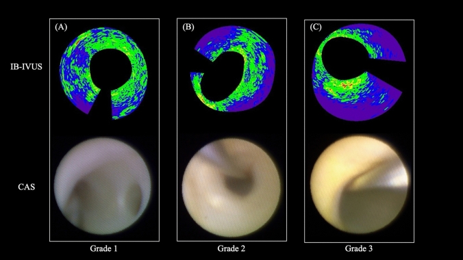
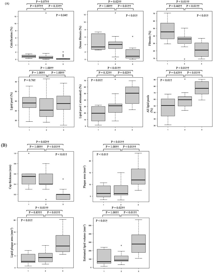
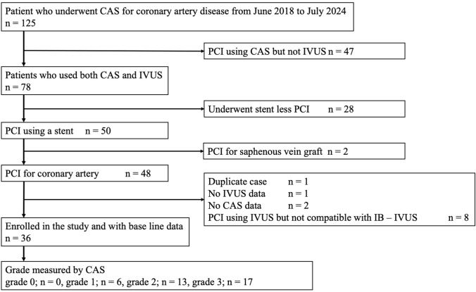
Coronary angioscopy (CAS) enables direct qualitative assessment of the coronary artery lumen, while integrated backscatter intravascular ultrasound (IB-IVUS) provides a quantitative evaluation of coronary plaque tissue characteristics. Despite the utility of both techniques in assessing coronary plaque status, data on the correlation between their findings remain limited. To investigate the association between CAS-derived findings and results obtained through IB-IVUS. This retrospective analysis included 36 patients who underwent both CAS and IB-IVUS during percutaneous coronary intervention (PCI) at our institution. CAS and IB-IVUS were performed on the same coronary artery treated during PCI. Plaques were categorized into four groups based on their yellow color grade using CAS. For the IB-IVUS analysis, measurements were performed at the minimum lumen diameter site of the culprit lesion. A significant correlation was observed between plaque yellowishness and plaque characteristics on IB-IVUS. Higher plaque yellowishness was associated with an increased percentage of all lipid pool (P < 0.01), a greater proportion of attenuated plaque (P < 0.01), and a larger estimated lipid plaque volume (P < 0.01). Additionally, plaques with higher yellowishness grades had significantly thinner fibrous caps (P < 0.01). The findings suggest that higher plaque yellowishness observed via CAS correlates with a larger lipid plaque volume and thinner fibrous caps, as assessed through IB-IVUS.
期刊介绍:
Cardiovascular Intervention and Therapeutics (CVIT) is an international journal covering the field of cardiovascular disease and includes cardiac (coronary and noncoronary) and peripheral interventions and therapeutics. Articles are subject to peer review and complete editorial evaluation prior to any decision regarding acceptability. CVIT is an official journal of The Japanese Association of Cardiovascular Intervention and Therapeutics.
 求助内容:
求助内容: 应助结果提醒方式:


