Sanket Dash, Sameer Vyas, Chirag Kamal Ahuja, Paramjeet Singh, Sarfraj Ahmad
{"title":"合成磁共振弛豫法鉴别中枢神经系统结核瘤和胶质母细胞瘤。","authors":"Sanket Dash, Sameer Vyas, Chirag Kamal Ahuja, Paramjeet Singh, Sarfraj Ahmad","doi":"10.5114/pjr/202175","DOIUrl":null,"url":null,"abstract":"<p><strong>Purpose: </strong>Synthetic magnetic resonance imaging (MRI) allows reconstruction of multiple contrast-weighted images from a single acquisition of multiple delay multiple echo (MDME) sequence with quantitative relaxometry (longitudinal relaxation rate [R1], transverse relaxation rate [R2], and proton density [PD]) in a shorter acquisition time. We tried to explore synthetic MR-based relaxometry to differentiate central nervous system (CNS) tuberculomas from primary CNS neoplasm like glioblastoma.</p><p><strong>Material and methods: </strong>Ten cases of CNS tuberculoma and 14 cases of glioblastoma underwent pre- and post-contrast synthetic MRI. R1, R2, and PD values were calculated from lesion core, wall, and perilesional oedema using free-hand region of interest and compared across the 2 groups.</p><p><strong>Results: </strong>Both pre- and post-contrast R1 and R2 relaxation rates from core were significantly higher in tuberculoma (mean pre-contrast R1 - 0.93, R2 - 15.02; post-contrast R1 - 1.51, R2 - 15.48) from glioblastoma (mean pre-contrast R1 - 0.36, R2 - 4.58; post-contrast R1 - 0.43, R2 - 4.78). The same values were higher in perilesional oedema of glioblastoma (mean pre-contrast R1 - 0.75, R2 - 9.9; post-contrast R1 - 0.78, R2 - 10.48) compared to tuberculoma (mean pre-contrast R1 - 0.68, R2 - 8.57; post-contrast R1 - 0.72, R2 - 8.67). No significant difference was seen between relaxometry parameters from the walls of lesions.</p><p><strong>Conclusions: </strong>Synthetic MR-based relaxometry can be useful in distinguishing CNS tuberculomas from glioblastoma. R1 and R2 relaxation rates from core of the lesions are most important in differentiating the two with R1 value > 0.852 and R2 value > 11.565 from core strongly suggests tuberculoma over glioblastoma.</p>","PeriodicalId":94174,"journal":{"name":"Polish journal of radiology","volume":"90 ","pages":"e198-e206"},"PeriodicalIF":0.0000,"publicationDate":"2025-04-29","publicationTypes":"Journal Article","fieldsOfStudy":null,"isOpenAccess":false,"openAccessPdf":"https://www.ncbi.nlm.nih.gov/pmc/articles/PMC12099202/pdf/","citationCount":"0","resultStr":"{\"title\":\"Synthetic magnetic resonance-based relaxometry in differentiating central nervous system tuberculoma and glioblastoma.\",\"authors\":\"Sanket Dash, Sameer Vyas, Chirag Kamal Ahuja, Paramjeet Singh, Sarfraj Ahmad\",\"doi\":\"10.5114/pjr/202175\",\"DOIUrl\":null,\"url\":null,\"abstract\":\"<p><strong>Purpose: </strong>Synthetic magnetic resonance imaging (MRI) allows reconstruction of multiple contrast-weighted images from a single acquisition of multiple delay multiple echo (MDME) sequence with quantitative relaxometry (longitudinal relaxation rate [R1], transverse relaxation rate [R2], and proton density [PD]) in a shorter acquisition time. We tried to explore synthetic MR-based relaxometry to differentiate central nervous system (CNS) tuberculomas from primary CNS neoplasm like glioblastoma.</p><p><strong>Material and methods: </strong>Ten cases of CNS tuberculoma and 14 cases of glioblastoma underwent pre- and post-contrast synthetic MRI. R1, R2, and PD values were calculated from lesion core, wall, and perilesional oedema using free-hand region of interest and compared across the 2 groups.</p><p><strong>Results: </strong>Both pre- and post-contrast R1 and R2 relaxation rates from core were significantly higher in tuberculoma (mean pre-contrast R1 - 0.93, R2 - 15.02; post-contrast R1 - 1.51, R2 - 15.48) from glioblastoma (mean pre-contrast R1 - 0.36, R2 - 4.58; post-contrast R1 - 0.43, R2 - 4.78). The same values were higher in perilesional oedema of glioblastoma (mean pre-contrast R1 - 0.75, R2 - 9.9; post-contrast R1 - 0.78, R2 - 10.48) compared to tuberculoma (mean pre-contrast R1 - 0.68, R2 - 8.57; post-contrast R1 - 0.72, R2 - 8.67). No significant difference was seen between relaxometry parameters from the walls of lesions.</p><p><strong>Conclusions: </strong>Synthetic MR-based relaxometry can be useful in distinguishing CNS tuberculomas from glioblastoma. R1 and R2 relaxation rates from core of the lesions are most important in differentiating the two with R1 value > 0.852 and R2 value > 11.565 from core strongly suggests tuberculoma over glioblastoma.</p>\",\"PeriodicalId\":94174,\"journal\":{\"name\":\"Polish journal of radiology\",\"volume\":\"90 \",\"pages\":\"e198-e206\"},\"PeriodicalIF\":0.0000,\"publicationDate\":\"2025-04-29\",\"publicationTypes\":\"Journal Article\",\"fieldsOfStudy\":null,\"isOpenAccess\":false,\"openAccessPdf\":\"https://www.ncbi.nlm.nih.gov/pmc/articles/PMC12099202/pdf/\",\"citationCount\":\"0\",\"resultStr\":null,\"platform\":\"Semanticscholar\",\"paperid\":null,\"PeriodicalName\":\"Polish journal of radiology\",\"FirstCategoryId\":\"1085\",\"ListUrlMain\":\"https://doi.org/10.5114/pjr/202175\",\"RegionNum\":0,\"RegionCategory\":null,\"ArticlePicture\":[],\"TitleCN\":null,\"AbstractTextCN\":null,\"PMCID\":null,\"EPubDate\":\"2025/1/1 0:00:00\",\"PubModel\":\"eCollection\",\"JCR\":\"\",\"JCRName\":\"\",\"Score\":null,\"Total\":0}","platform":"Semanticscholar","paperid":null,"PeriodicalName":"Polish journal of radiology","FirstCategoryId":"1085","ListUrlMain":"https://doi.org/10.5114/pjr/202175","RegionNum":0,"RegionCategory":null,"ArticlePicture":[],"TitleCN":null,"AbstractTextCN":null,"PMCID":null,"EPubDate":"2025/1/1 0:00:00","PubModel":"eCollection","JCR":"","JCRName":"","Score":null,"Total":0}
Synthetic magnetic resonance-based relaxometry in differentiating central nervous system tuberculoma and glioblastoma.
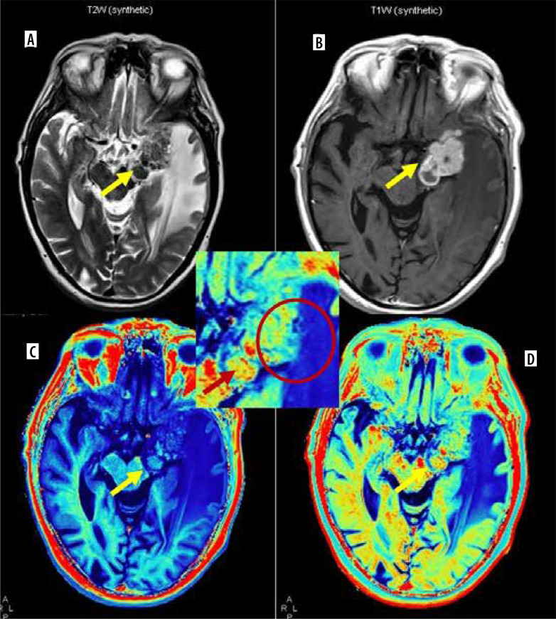
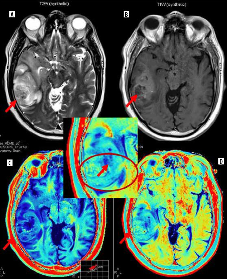
Purpose: Synthetic magnetic resonance imaging (MRI) allows reconstruction of multiple contrast-weighted images from a single acquisition of multiple delay multiple echo (MDME) sequence with quantitative relaxometry (longitudinal relaxation rate [R1], transverse relaxation rate [R2], and proton density [PD]) in a shorter acquisition time. We tried to explore synthetic MR-based relaxometry to differentiate central nervous system (CNS) tuberculomas from primary CNS neoplasm like glioblastoma.
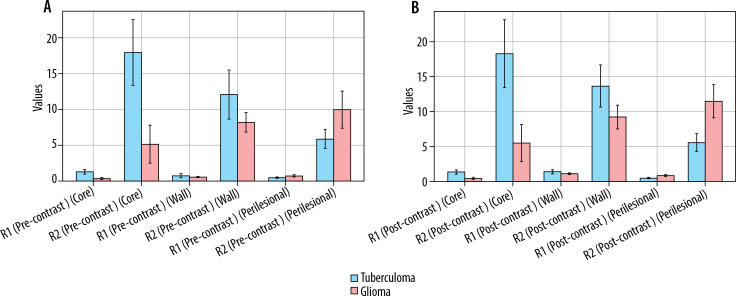
Material and methods: Ten cases of CNS tuberculoma and 14 cases of glioblastoma underwent pre- and post-contrast synthetic MRI. R1, R2, and PD values were calculated from lesion core, wall, and perilesional oedema using free-hand region of interest and compared across the 2 groups.
Results: Both pre- and post-contrast R1 and R2 relaxation rates from core were significantly higher in tuberculoma (mean pre-contrast R1 - 0.93, R2 - 15.02; post-contrast R1 - 1.51, R2 - 15.48) from glioblastoma (mean pre-contrast R1 - 0.36, R2 - 4.58; post-contrast R1 - 0.43, R2 - 4.78). The same values were higher in perilesional oedema of glioblastoma (mean pre-contrast R1 - 0.75, R2 - 9.9; post-contrast R1 - 0.78, R2 - 10.48) compared to tuberculoma (mean pre-contrast R1 - 0.68, R2 - 8.57; post-contrast R1 - 0.72, R2 - 8.67). No significant difference was seen between relaxometry parameters from the walls of lesions.
Conclusions: Synthetic MR-based relaxometry can be useful in distinguishing CNS tuberculomas from glioblastoma. R1 and R2 relaxation rates from core of the lesions are most important in differentiating the two with R1 value > 0.852 and R2 value > 11.565 from core strongly suggests tuberculoma over glioblastoma.

 求助内容:
求助内容: 应助结果提醒方式:


