Soon Chul Heo, Hae Won Shin, Dong Joon Lee, Franklin Garcia-Godoy, Bo Ram Keum, Yong Hoon Kwon, Hyung Joon Kim
{"title":"酒石酸支链聚乙烯亚胺碳点通过成骨分化促进骨缺损修复。","authors":"Soon Chul Heo, Hae Won Shin, Dong Joon Lee, Franklin Garcia-Godoy, Bo Ram Keum, Yong Hoon Kwon, Hyung Joon Kim","doi":"10.1093/rb/rbaf030","DOIUrl":null,"url":null,"abstract":"<p><p>Treating bone defects is a critical challenge in regenerative medicine. Carbon nanomaterials, with their unique physicochemical properties, offer significant potential for enhancing bone regeneration. In this study, we developed tartaric acid (TA)-based carbon dots (CDs) by synthesizing TA with branched polyethyleneimine (bPEI). These TA-bPEI CDs were systematically evaluated to determine their effects on osteogenic differentiation in human bone marrow-derived mesenchymal stem cells (BMSCs) and their capacity to repair calvarial defects in an <i>in vivo</i> model. Characterization of TA-bPEI CDs revealed a size of approximately 10 nm and a positive surface charge. The CDs exhibited fluorescence emission peaks between 464 and 506 nm under excitation wavelengths of 340-440 nm. Cytotoxicity assays demonstrated that TA-bPEI CDs maintained BMSC viability at concentrations up to 250 μg/ml. However, at concentrations of 500 μg/ml and above, apoptosis was induced. Treatment with TA-bPEI significantly enhanced osteogenic differentiation <i>in vitro</i>, as evidenced by increased expression of osteogenic-specific proteins such as Runx2, ALP, OCN and OPN. <i>In vivo</i>, the application of TA-bPEI CDs in a mouse calvarial defect model promoted robust new bone formation, reduced defect gaps, and improved bone morphometric parameters, including bone volume fraction and trabecular thickness. These results suggest that TA-bPEI CDs enhance osteogenesis by directly stimulating osteogenic differentiation and upregulating osteogenesis-specific genes. This study demonstrates the high potential of TA-bPEI CDs as a novel nanomaterial for bone regeneration applications.</p>","PeriodicalId":20929,"journal":{"name":"Regenerative Biomaterials","volume":"12 ","pages":"rbaf030"},"PeriodicalIF":8.1000,"publicationDate":"2025-05-16","publicationTypes":"Journal Article","fieldsOfStudy":null,"isOpenAccess":false,"openAccessPdf":"https://www.ncbi.nlm.nih.gov/pmc/articles/PMC12098262/pdf/","citationCount":"0","resultStr":"{\"title\":\"Tartaric acid-branched polyethyleneimine carbon dots promote repair of bone defect via osteogenic differentiation.\",\"authors\":\"Soon Chul Heo, Hae Won Shin, Dong Joon Lee, Franklin Garcia-Godoy, Bo Ram Keum, Yong Hoon Kwon, Hyung Joon Kim\",\"doi\":\"10.1093/rb/rbaf030\",\"DOIUrl\":null,\"url\":null,\"abstract\":\"<p><p>Treating bone defects is a critical challenge in regenerative medicine. Carbon nanomaterials, with their unique physicochemical properties, offer significant potential for enhancing bone regeneration. In this study, we developed tartaric acid (TA)-based carbon dots (CDs) by synthesizing TA with branched polyethyleneimine (bPEI). These TA-bPEI CDs were systematically evaluated to determine their effects on osteogenic differentiation in human bone marrow-derived mesenchymal stem cells (BMSCs) and their capacity to repair calvarial defects in an <i>in vivo</i> model. Characterization of TA-bPEI CDs revealed a size of approximately 10 nm and a positive surface charge. The CDs exhibited fluorescence emission peaks between 464 and 506 nm under excitation wavelengths of 340-440 nm. Cytotoxicity assays demonstrated that TA-bPEI CDs maintained BMSC viability at concentrations up to 250 μg/ml. However, at concentrations of 500 μg/ml and above, apoptosis was induced. Treatment with TA-bPEI significantly enhanced osteogenic differentiation <i>in vitro</i>, as evidenced by increased expression of osteogenic-specific proteins such as Runx2, ALP, OCN and OPN. <i>In vivo</i>, the application of TA-bPEI CDs in a mouse calvarial defect model promoted robust new bone formation, reduced defect gaps, and improved bone morphometric parameters, including bone volume fraction and trabecular thickness. These results suggest that TA-bPEI CDs enhance osteogenesis by directly stimulating osteogenic differentiation and upregulating osteogenesis-specific genes. This study demonstrates the high potential of TA-bPEI CDs as a novel nanomaterial for bone regeneration applications.</p>\",\"PeriodicalId\":20929,\"journal\":{\"name\":\"Regenerative Biomaterials\",\"volume\":\"12 \",\"pages\":\"rbaf030\"},\"PeriodicalIF\":8.1000,\"publicationDate\":\"2025-05-16\",\"publicationTypes\":\"Journal Article\",\"fieldsOfStudy\":null,\"isOpenAccess\":false,\"openAccessPdf\":\"https://www.ncbi.nlm.nih.gov/pmc/articles/PMC12098262/pdf/\",\"citationCount\":\"0\",\"resultStr\":null,\"platform\":\"Semanticscholar\",\"paperid\":null,\"PeriodicalName\":\"Regenerative Biomaterials\",\"FirstCategoryId\":\"5\",\"ListUrlMain\":\"https://doi.org/10.1093/rb/rbaf030\",\"RegionNum\":1,\"RegionCategory\":\"医学\",\"ArticlePicture\":[],\"TitleCN\":null,\"AbstractTextCN\":null,\"PMCID\":null,\"EPubDate\":\"2025/1/1 0:00:00\",\"PubModel\":\"eCollection\",\"JCR\":\"Q1\",\"JCRName\":\"MATERIALS SCIENCE, BIOMATERIALS\",\"Score\":null,\"Total\":0}","platform":"Semanticscholar","paperid":null,"PeriodicalName":"Regenerative Biomaterials","FirstCategoryId":"5","ListUrlMain":"https://doi.org/10.1093/rb/rbaf030","RegionNum":1,"RegionCategory":"医学","ArticlePicture":[],"TitleCN":null,"AbstractTextCN":null,"PMCID":null,"EPubDate":"2025/1/1 0:00:00","PubModel":"eCollection","JCR":"Q1","JCRName":"MATERIALS SCIENCE, BIOMATERIALS","Score":null,"Total":0}
Tartaric acid-branched polyethyleneimine carbon dots promote repair of bone defect via osteogenic differentiation.
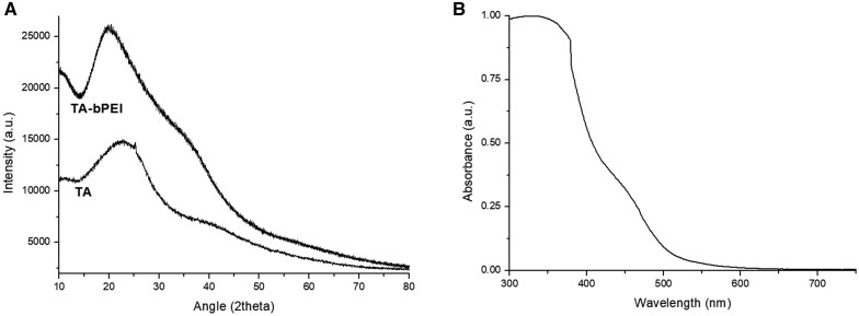
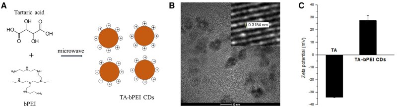
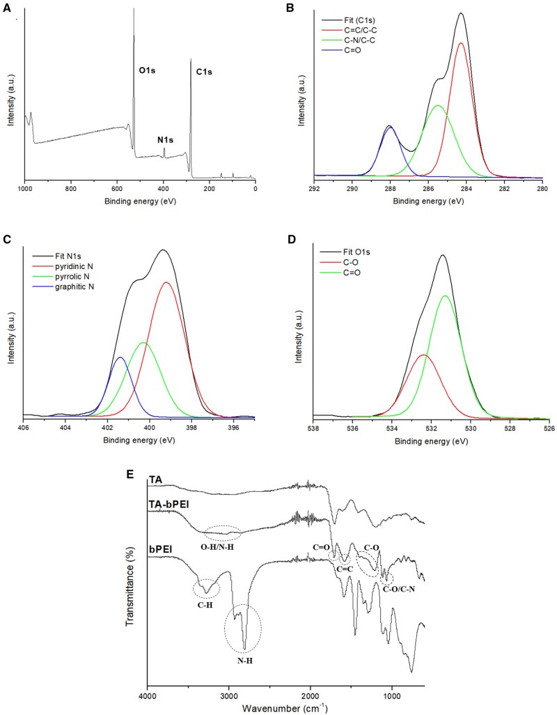
Treating bone defects is a critical challenge in regenerative medicine. Carbon nanomaterials, with their unique physicochemical properties, offer significant potential for enhancing bone regeneration. In this study, we developed tartaric acid (TA)-based carbon dots (CDs) by synthesizing TA with branched polyethyleneimine (bPEI). These TA-bPEI CDs were systematically evaluated to determine their effects on osteogenic differentiation in human bone marrow-derived mesenchymal stem cells (BMSCs) and their capacity to repair calvarial defects in an in vivo model. Characterization of TA-bPEI CDs revealed a size of approximately 10 nm and a positive surface charge. The CDs exhibited fluorescence emission peaks between 464 and 506 nm under excitation wavelengths of 340-440 nm. Cytotoxicity assays demonstrated that TA-bPEI CDs maintained BMSC viability at concentrations up to 250 μg/ml. However, at concentrations of 500 μg/ml and above, apoptosis was induced. Treatment with TA-bPEI significantly enhanced osteogenic differentiation in vitro, as evidenced by increased expression of osteogenic-specific proteins such as Runx2, ALP, OCN and OPN. In vivo, the application of TA-bPEI CDs in a mouse calvarial defect model promoted robust new bone formation, reduced defect gaps, and improved bone morphometric parameters, including bone volume fraction and trabecular thickness. These results suggest that TA-bPEI CDs enhance osteogenesis by directly stimulating osteogenic differentiation and upregulating osteogenesis-specific genes. This study demonstrates the high potential of TA-bPEI CDs as a novel nanomaterial for bone regeneration applications.
期刊介绍:
Regenerative Biomaterials is an international, interdisciplinary, peer-reviewed journal publishing the latest advances in biomaterials and regenerative medicine. The journal provides a forum for the publication of original research papers, reviews, clinical case reports, and commentaries on the topics relevant to the development of advanced regenerative biomaterials concerning novel regenerative technologies and therapeutic approaches for the regeneration and repair of damaged tissues and organs. The interactions of biomaterials with cells and tissue, especially with stem cells, will be of particular focus.
 求助内容:
求助内容: 应助结果提醒方式:


