Stella Den Hengst, Noor Borren, Esther M M Van Lieshout, Job N Doornberg, Theo Van Walsum, Mathieu M E Wijffels, Michael H J Verhofstad
{"title":"利用深度学习模型从CT数据中检测、分类和分割肋骨骨折:文献综述和汇总分析。","authors":"Stella Den Hengst, Noor Borren, Esther M M Van Lieshout, Job N Doornberg, Theo Van Walsum, Mathieu M E Wijffels, Michael H J Verhofstad","doi":"10.1097/RTI.0000000000000833","DOIUrl":null,"url":null,"abstract":"<p><strong>Purpose: </strong>Trauma-induced rib fractures are common injuries. The gold standard for diagnosing rib fractures is computed tomography (CT), but the sensitivity in the acute setting is low, and interpreting CT slices is labor-intensive. This has led to the development of new diagnostic approaches leveraging deep learning (DL) models. This systematic review and pooled analysis aimed to compare the performance of DL models in the detection, segmentation, and classification of rib fractures based on CT scans.</p><p><strong>Materials and methods: </strong>A literature search was performed using various databases for studies describing DL models detecting, segmenting, or classifying rib fractures from CT data. Reported performance metrics included sensitivity, false-positive rate, F1-score, precision, accuracy, and mean average precision. A meta-analysis was performed on the sensitivity scores to compare the DL models with clinicians.</p><p><strong>Results: </strong>Of the 323 identified records, 25 were included. Twenty-one studies reported on detection, four on segmentation, and 10 on classification. Twenty studies had adequate data for meta-analysis. The gold standard labels were provided by clinicians who were radiologists and orthopedic surgeons. For detecting rib fractures, DL models had a higher sensitivity (86.7%; 95% CI: 82.6%-90.2%) than clinicians (75.4%; 95% CI: 68.1%-82.1%). In classification, the sensitivity of DL models for displaced rib fractures (97.3%; 95% CI: 95.6%-98.5%) was significantly better than that of clinicians (88.2%; 95% CI: 84.8%-91.3%).</p><p><strong>Conclusions: </strong>DL models for rib fracture detection and classification achieved promising results. With better sensitivities than clinicians for detecting and classifying displaced rib fractures, the future should focus on implementing DL models in daily clinics.</p><p><strong>Level of evidence: </strong>Level III-systematic review and pooled analysis.</p>","PeriodicalId":49974,"journal":{"name":"Journal of Thoracic Imaging","volume":" ","pages":""},"PeriodicalIF":1.9000,"publicationDate":"2025-09-01","publicationTypes":"Journal Article","fieldsOfStudy":null,"isOpenAccess":false,"openAccessPdf":"https://www.ncbi.nlm.nih.gov/pmc/articles/PMC12369507/pdf/","citationCount":"0","resultStr":"{\"title\":\"Detection, Classification, and Segmentation of Rib Fractures From CT Data Using Deep Learning Models: A Review of Literature and Pooled Analysis.\",\"authors\":\"Stella Den Hengst, Noor Borren, Esther M M Van Lieshout, Job N Doornberg, Theo Van Walsum, Mathieu M E Wijffels, Michael H J Verhofstad\",\"doi\":\"10.1097/RTI.0000000000000833\",\"DOIUrl\":null,\"url\":null,\"abstract\":\"<p><strong>Purpose: </strong>Trauma-induced rib fractures are common injuries. The gold standard for diagnosing rib fractures is computed tomography (CT), but the sensitivity in the acute setting is low, and interpreting CT slices is labor-intensive. This has led to the development of new diagnostic approaches leveraging deep learning (DL) models. This systematic review and pooled analysis aimed to compare the performance of DL models in the detection, segmentation, and classification of rib fractures based on CT scans.</p><p><strong>Materials and methods: </strong>A literature search was performed using various databases for studies describing DL models detecting, segmenting, or classifying rib fractures from CT data. Reported performance metrics included sensitivity, false-positive rate, F1-score, precision, accuracy, and mean average precision. A meta-analysis was performed on the sensitivity scores to compare the DL models with clinicians.</p><p><strong>Results: </strong>Of the 323 identified records, 25 were included. Twenty-one studies reported on detection, four on segmentation, and 10 on classification. Twenty studies had adequate data for meta-analysis. The gold standard labels were provided by clinicians who were radiologists and orthopedic surgeons. For detecting rib fractures, DL models had a higher sensitivity (86.7%; 95% CI: 82.6%-90.2%) than clinicians (75.4%; 95% CI: 68.1%-82.1%). In classification, the sensitivity of DL models for displaced rib fractures (97.3%; 95% CI: 95.6%-98.5%) was significantly better than that of clinicians (88.2%; 95% CI: 84.8%-91.3%).</p><p><strong>Conclusions: </strong>DL models for rib fracture detection and classification achieved promising results. With better sensitivities than clinicians for detecting and classifying displaced rib fractures, the future should focus on implementing DL models in daily clinics.</p><p><strong>Level of evidence: </strong>Level III-systematic review and pooled analysis.</p>\",\"PeriodicalId\":49974,\"journal\":{\"name\":\"Journal of Thoracic Imaging\",\"volume\":\" \",\"pages\":\"\"},\"PeriodicalIF\":1.9000,\"publicationDate\":\"2025-09-01\",\"publicationTypes\":\"Journal Article\",\"fieldsOfStudy\":null,\"isOpenAccess\":false,\"openAccessPdf\":\"https://www.ncbi.nlm.nih.gov/pmc/articles/PMC12369507/pdf/\",\"citationCount\":\"0\",\"resultStr\":null,\"platform\":\"Semanticscholar\",\"paperid\":null,\"PeriodicalName\":\"Journal of Thoracic Imaging\",\"FirstCategoryId\":\"3\",\"ListUrlMain\":\"https://doi.org/10.1097/RTI.0000000000000833\",\"RegionNum\":4,\"RegionCategory\":\"医学\",\"ArticlePicture\":[],\"TitleCN\":null,\"AbstractTextCN\":null,\"PMCID\":null,\"EPubDate\":\"\",\"PubModel\":\"\",\"JCR\":\"Q3\",\"JCRName\":\"RADIOLOGY, NUCLEAR MEDICINE & MEDICAL IMAGING\",\"Score\":null,\"Total\":0}","platform":"Semanticscholar","paperid":null,"PeriodicalName":"Journal of Thoracic Imaging","FirstCategoryId":"3","ListUrlMain":"https://doi.org/10.1097/RTI.0000000000000833","RegionNum":4,"RegionCategory":"医学","ArticlePicture":[],"TitleCN":null,"AbstractTextCN":null,"PMCID":null,"EPubDate":"","PubModel":"","JCR":"Q3","JCRName":"RADIOLOGY, NUCLEAR MEDICINE & MEDICAL IMAGING","Score":null,"Total":0}
Detection, Classification, and Segmentation of Rib Fractures From CT Data Using Deep Learning Models: A Review of Literature and Pooled Analysis.
Purpose: Trauma-induced rib fractures are common injuries. The gold standard for diagnosing rib fractures is computed tomography (CT), but the sensitivity in the acute setting is low, and interpreting CT slices is labor-intensive. This has led to the development of new diagnostic approaches leveraging deep learning (DL) models. This systematic review and pooled analysis aimed to compare the performance of DL models in the detection, segmentation, and classification of rib fractures based on CT scans.
Materials and methods: A literature search was performed using various databases for studies describing DL models detecting, segmenting, or classifying rib fractures from CT data. Reported performance metrics included sensitivity, false-positive rate, F1-score, precision, accuracy, and mean average precision. A meta-analysis was performed on the sensitivity scores to compare the DL models with clinicians.
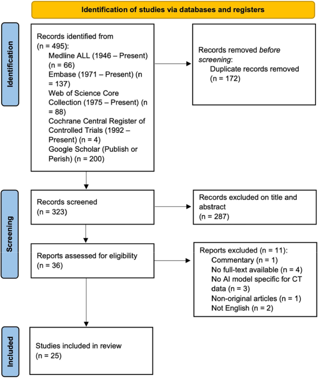
Results: Of the 323 identified records, 25 were included. Twenty-one studies reported on detection, four on segmentation, and 10 on classification. Twenty studies had adequate data for meta-analysis. The gold standard labels were provided by clinicians who were radiologists and orthopedic surgeons. For detecting rib fractures, DL models had a higher sensitivity (86.7%; 95% CI: 82.6%-90.2%) than clinicians (75.4%; 95% CI: 68.1%-82.1%). In classification, the sensitivity of DL models for displaced rib fractures (97.3%; 95% CI: 95.6%-98.5%) was significantly better than that of clinicians (88.2%; 95% CI: 84.8%-91.3%).
Conclusions: DL models for rib fracture detection and classification achieved promising results. With better sensitivities than clinicians for detecting and classifying displaced rib fractures, the future should focus on implementing DL models in daily clinics.
Level of evidence: Level III-systematic review and pooled analysis.
期刊介绍:
Journal of Thoracic Imaging (JTI) provides authoritative information on all aspects of the use of imaging techniques in the diagnosis of cardiac and pulmonary diseases. Original articles and analytical reviews published in this timely journal provide the very latest thinking of leading experts concerning the use of chest radiography, computed tomography, magnetic resonance imaging, positron emission tomography, ultrasound, and all other promising imaging techniques in cardiopulmonary radiology.
Official Journal of the Society of Thoracic Radiology:
Japanese Society of Thoracic Radiology
Korean Society of Thoracic Radiology
European Society of Thoracic Imaging.

 求助内容:
求助内容: 应助结果提醒方式:


