Manuel R Pouso, Emanuel Farinha, Henrique E Costa, Margarida Lorigo, Graça Baltazar, Elisa Cairrao
{"title":"维甲酸对脑血管系统的影响:正常和缺血情况下平滑肌细胞血管活性反应的分析。","authors":"Manuel R Pouso, Emanuel Farinha, Henrique E Costa, Margarida Lorigo, Graça Baltazar, Elisa Cairrao","doi":"10.3390/jox15030069","DOIUrl":null,"url":null,"abstract":"<p><p>Retinoic acid (RA), a vitamin A derivative, has been shown to prevent the development of neurological disorders by ensuring the integrity of the physiological structure of the neurovascular unit and regulating the physiological cell's function. After an ischemia event, RA reduces the effects of blood-brain barrier disruption by blocking the apoptotic signaling pathway. However, the effect of RA on smooth muscle cells (SMCs), which are crucial to maintaining blood perfusion, has never been investigated. This study aimed to evaluate the effect of RA on the vasoactive response of middle cerebral artery SMCs in normal and ischemic contexts (O<sub>2</sub> and glucose deprivation, OGD). For this purpose, SMCs cultures were incubated with RA, and the vasoactive response was evaluated in both conditions (OGD and non-OGD). To simulate OGD, co-cultures of neurons and astrocytes were made and incubated with RA to analyze the effect of the secretome released by these cells on SMCs contractility. In non-OGD conditions, RA induced rapid relaxation of SMCs and, in the long term (24 h), promoted cell contraction. In OGD conditions, SMCs contractility patterns were different when pre-incubated with RA. In these conditions, NA loses its contractility effect, and SNP seems to revert its relaxant effect. However, SMCs pre-incubated with 5 uM RA show the vasorelaxant pattern typical of SNP, despite the OGD condition. These effects demonstrate an effect of RA on the vasoactive profile of SMCs, with therapeutic potential in OGD conditions.</p>","PeriodicalId":42356,"journal":{"name":"Journal of Xenobiotics","volume":"15 3","pages":""},"PeriodicalIF":4.4000,"publicationDate":"2025-05-10","publicationTypes":"Journal Article","fieldsOfStudy":null,"isOpenAccess":false,"openAccessPdf":"https://www.ncbi.nlm.nih.gov/pmc/articles/PMC12101329/pdf/","citationCount":"0","resultStr":"{\"title\":\"Effect of Retinoic Acid on the Cerebral Vasculature: Analysis of the Vasoactive Response of Smooth Muscle Cells in Normal and Ischemic Contexts.\",\"authors\":\"Manuel R Pouso, Emanuel Farinha, Henrique E Costa, Margarida Lorigo, Graça Baltazar, Elisa Cairrao\",\"doi\":\"10.3390/jox15030069\",\"DOIUrl\":null,\"url\":null,\"abstract\":\"<p><p>Retinoic acid (RA), a vitamin A derivative, has been shown to prevent the development of neurological disorders by ensuring the integrity of the physiological structure of the neurovascular unit and regulating the physiological cell's function. After an ischemia event, RA reduces the effects of blood-brain barrier disruption by blocking the apoptotic signaling pathway. However, the effect of RA on smooth muscle cells (SMCs), which are crucial to maintaining blood perfusion, has never been investigated. This study aimed to evaluate the effect of RA on the vasoactive response of middle cerebral artery SMCs in normal and ischemic contexts (O<sub>2</sub> and glucose deprivation, OGD). For this purpose, SMCs cultures were incubated with RA, and the vasoactive response was evaluated in both conditions (OGD and non-OGD). To simulate OGD, co-cultures of neurons and astrocytes were made and incubated with RA to analyze the effect of the secretome released by these cells on SMCs contractility. In non-OGD conditions, RA induced rapid relaxation of SMCs and, in the long term (24 h), promoted cell contraction. In OGD conditions, SMCs contractility patterns were different when pre-incubated with RA. In these conditions, NA loses its contractility effect, and SNP seems to revert its relaxant effect. However, SMCs pre-incubated with 5 uM RA show the vasorelaxant pattern typical of SNP, despite the OGD condition. These effects demonstrate an effect of RA on the vasoactive profile of SMCs, with therapeutic potential in OGD conditions.</p>\",\"PeriodicalId\":42356,\"journal\":{\"name\":\"Journal of Xenobiotics\",\"volume\":\"15 3\",\"pages\":\"\"},\"PeriodicalIF\":4.4000,\"publicationDate\":\"2025-05-10\",\"publicationTypes\":\"Journal Article\",\"fieldsOfStudy\":null,\"isOpenAccess\":false,\"openAccessPdf\":\"https://www.ncbi.nlm.nih.gov/pmc/articles/PMC12101329/pdf/\",\"citationCount\":\"0\",\"resultStr\":null,\"platform\":\"Semanticscholar\",\"paperid\":null,\"PeriodicalName\":\"Journal of Xenobiotics\",\"FirstCategoryId\":\"1085\",\"ListUrlMain\":\"https://doi.org/10.3390/jox15030069\",\"RegionNum\":0,\"RegionCategory\":null,\"ArticlePicture\":[],\"TitleCN\":null,\"AbstractTextCN\":null,\"PMCID\":null,\"EPubDate\":\"\",\"PubModel\":\"\",\"JCR\":\"Q1\",\"JCRName\":\"TOXICOLOGY\",\"Score\":null,\"Total\":0}","platform":"Semanticscholar","paperid":null,"PeriodicalName":"Journal of Xenobiotics","FirstCategoryId":"1085","ListUrlMain":"https://doi.org/10.3390/jox15030069","RegionNum":0,"RegionCategory":null,"ArticlePicture":[],"TitleCN":null,"AbstractTextCN":null,"PMCID":null,"EPubDate":"","PubModel":"","JCR":"Q1","JCRName":"TOXICOLOGY","Score":null,"Total":0}
引用次数: 0
摘要
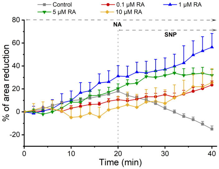
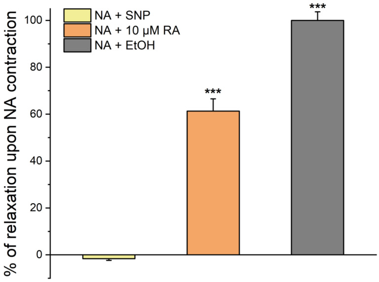
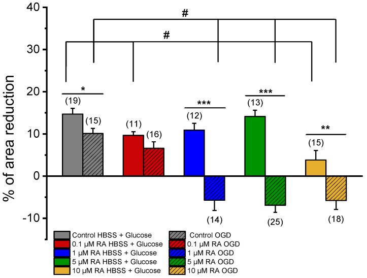
维甲酸(RA)是维生素a的衍生物,已被证明可以通过确保神经血管单位生理结构的完整性和调节生理细胞的功能来预防神经系统疾病的发展。在缺血事件后,RA通过阻断凋亡信号通路减少血脑屏障破坏的影响。然而,RA对维持血液灌注至关重要的平滑肌细胞(SMCs)的影响从未被研究过。本研究旨在评估RA对正常和缺血情况下大脑中动脉SMCs血管活性反应的影响(O2和glucose deprivation, OGD)。为此,SMCs培养物与RA一起孵育,并在两种情况下(OGD和非OGD)评估血管活性反应。为了模拟OGD,我们制作神经元和星形胶质细胞共培养,并与RA孵育,分析这些细胞释放的分泌组对SMCs收缩性的影响。在非ogd条件下,RA诱导SMCs快速松弛,并在长期(24小时)内促进细胞收缩。在OGD条件下,与RA预孵育的SMCs收缩模式不同。在这些情况下,NA失去了它的收缩作用,而SNP似乎恢复了它的松弛作用。然而,5 uM RA预孵育的SMCs显示SNP典型的血管松弛模式,尽管存在OGD情况。这些效应表明RA对SMCs的血管活性谱有影响,具有治疗OGD的潜力。
Effect of Retinoic Acid on the Cerebral Vasculature: Analysis of the Vasoactive Response of Smooth Muscle Cells in Normal and Ischemic Contexts.
Retinoic acid (RA), a vitamin A derivative, has been shown to prevent the development of neurological disorders by ensuring the integrity of the physiological structure of the neurovascular unit and regulating the physiological cell's function. After an ischemia event, RA reduces the effects of blood-brain barrier disruption by blocking the apoptotic signaling pathway. However, the effect of RA on smooth muscle cells (SMCs), which are crucial to maintaining blood perfusion, has never been investigated. This study aimed to evaluate the effect of RA on the vasoactive response of middle cerebral artery SMCs in normal and ischemic contexts (O2 and glucose deprivation, OGD). For this purpose, SMCs cultures were incubated with RA, and the vasoactive response was evaluated in both conditions (OGD and non-OGD). To simulate OGD, co-cultures of neurons and astrocytes were made and incubated with RA to analyze the effect of the secretome released by these cells on SMCs contractility. In non-OGD conditions, RA induced rapid relaxation of SMCs and, in the long term (24 h), promoted cell contraction. In OGD conditions, SMCs contractility patterns were different when pre-incubated with RA. In these conditions, NA loses its contractility effect, and SNP seems to revert its relaxant effect. However, SMCs pre-incubated with 5 uM RA show the vasorelaxant pattern typical of SNP, despite the OGD condition. These effects demonstrate an effect of RA on the vasoactive profile of SMCs, with therapeutic potential in OGD conditions.
期刊介绍:
The Journal of Xenobiotics publishes original studies concerning the beneficial (pharmacology) and detrimental effects (toxicology) of xenobiotics in all organisms. A xenobiotic (“stranger to life”) is defined as a chemical that is not usually found at significant concentrations or expected to reside for long periods in organisms. In addition to man-made chemicals, natural products could also be of interest if they have potent biological properties, special medicinal properties or that a given organism is at risk of exposure in the environment. Topics dealing with abiotic- and biotic-based transformations in various media (xenobiochemistry) and environmental toxicology are also of interest. Areas of interests include the identification of key physical and chemical properties of molecules that predict biological effects and persistence in the environment; the molecular mode of action of xenobiotics; biochemical and physiological interactions leading to change in organism health; pathophysiological interactions of natural and synthetic chemicals; development of biochemical indicators including new “-omics” approaches to identify biomarkers of exposure or effects for xenobiotics.

 求助内容:
求助内容: 应助结果提醒方式:


