{"title":"高糖和羧甲基赖氨酸对骨细胞基因表达的影响。","authors":"Rachana Vaidya, Lauren Conlon, Olivia Duclos, Ramina Behzad, Jacob Aaronson, Lamya Karim","doi":"10.4236/ajmb.2025.152012","DOIUrl":null,"url":null,"abstract":"<p><p>Diabetes mellitus (DM) is associated with increased bone fragility despite normal or elevated bone mineral density, partially due to the accumulation of advanced glycation end products (AGEs) in bone tissue. AGEs, such as carboxymethyl lysine (CML), impair osteocyte function by activating the receptor for advanced glycation end products (RAGE), triggering oxidative stress and inflammatory responses. This study aimed to investigate the effects of high glucose (HG) and CML on bone remodeling, glycation, inflammatory markers, and cellular functions in osteocytes. Using the murine osteocyte cell line OCY454-12H, we treated cells with HG (30 mM glucose) or 3 μM CML to simulate diabetic conditions. We assessed the expression of bone remodeling markers (SOST, RANKL, OPG, CTsK), glycation markers (RAGE, AGER1), inflammatory cytokines (IL-6, TNF-<i>α</i>), and cellular functions, including proliferation, viability, and apoptosis, using quantitative PCR and functional assays. HG treatment resulted in a 10-fold increase in SOST expression (9.3 vs. 0.9, p ≤ 0.0001) and a 2.4-fold increase in RANKL expression (2.75 vs. 1.15, p ≤ 0.0001), with a concurrent 2-fold increase in OPG (2.60 vs. 1.04, p ≤ 0.0001). The RANKL/OPG ratio remained unchanged (p = 0.15). HG also significantly increased RAGE expression by 3.67-fold (4.20 vs. 1.15, p ≤ 0.0001) and AGER1 by 1.65-fold (1.94 vs. 1.15, p ≤ 0.0001), along with a 2.02-fold increase in IL-6 (2.32 vs. 1.12, p ≤ 0.001) and a 7.35-fold increase in TNF-<i>α</i> (7.04 vs. 1.04, p ≤ 0.0001). Cell viability and proliferation were significantly higher under HG, accompanied by increased caspase-3 activity, indicating enhanced apoptosis. In contrast, CML exposure significantly upregulated RAGE (3.18 vs. 1.15, p ≤ 0.0001) and AGER1 (2.10 vs. 1.14, p = 0.028) but had no significant effects on bone remodeling markers, inflammatory cytokines, or cellular functions at physiological concentrations. Our findings demonstrate that HG disrupts osteocyte function by altering bone remodeling, glycation, and inflammatory pathways, while CML at physiological levels selectively activates glycation markers without inducing broader cellular dysfunction. These results underscore the role of the AGE-RAGE axis in diabetic bone fragility and highlight the need for future <i>in vivo</i> studies to explore therapeutic strategies targeting AGE accumulation and RAGE signaling in bone.</p>","PeriodicalId":65391,"journal":{"name":"美国分子生物学期刊(英文)","volume":"15 2","pages":"150-169"},"PeriodicalIF":0.0000,"publicationDate":"2025-04-01","publicationTypes":"Journal Article","fieldsOfStudy":null,"isOpenAccess":false,"openAccessPdf":"https://www.ncbi.nlm.nih.gov/pmc/articles/PMC12094516/pdf/","citationCount":"0","resultStr":"{\"title\":\"Effect of High Glucose and Carboxymethyl-Lysine on Osteocyte Gene Expression.\",\"authors\":\"Rachana Vaidya, Lauren Conlon, Olivia Duclos, Ramina Behzad, Jacob Aaronson, Lamya Karim\",\"doi\":\"10.4236/ajmb.2025.152012\",\"DOIUrl\":null,\"url\":null,\"abstract\":\"<p><p>Diabetes mellitus (DM) is associated with increased bone fragility despite normal or elevated bone mineral density, partially due to the accumulation of advanced glycation end products (AGEs) in bone tissue. AGEs, such as carboxymethyl lysine (CML), impair osteocyte function by activating the receptor for advanced glycation end products (RAGE), triggering oxidative stress and inflammatory responses. This study aimed to investigate the effects of high glucose (HG) and CML on bone remodeling, glycation, inflammatory markers, and cellular functions in osteocytes. Using the murine osteocyte cell line OCY454-12H, we treated cells with HG (30 mM glucose) or 3 μM CML to simulate diabetic conditions. We assessed the expression of bone remodeling markers (SOST, RANKL, OPG, CTsK), glycation markers (RAGE, AGER1), inflammatory cytokines (IL-6, TNF-<i>α</i>), and cellular functions, including proliferation, viability, and apoptosis, using quantitative PCR and functional assays. HG treatment resulted in a 10-fold increase in SOST expression (9.3 vs. 0.9, p ≤ 0.0001) and a 2.4-fold increase in RANKL expression (2.75 vs. 1.15, p ≤ 0.0001), with a concurrent 2-fold increase in OPG (2.60 vs. 1.04, p ≤ 0.0001). The RANKL/OPG ratio remained unchanged (p = 0.15). HG also significantly increased RAGE expression by 3.67-fold (4.20 vs. 1.15, p ≤ 0.0001) and AGER1 by 1.65-fold (1.94 vs. 1.15, p ≤ 0.0001), along with a 2.02-fold increase in IL-6 (2.32 vs. 1.12, p ≤ 0.001) and a 7.35-fold increase in TNF-<i>α</i> (7.04 vs. 1.04, p ≤ 0.0001). Cell viability and proliferation were significantly higher under HG, accompanied by increased caspase-3 activity, indicating enhanced apoptosis. In contrast, CML exposure significantly upregulated RAGE (3.18 vs. 1.15, p ≤ 0.0001) and AGER1 (2.10 vs. 1.14, p = 0.028) but had no significant effects on bone remodeling markers, inflammatory cytokines, or cellular functions at physiological concentrations. Our findings demonstrate that HG disrupts osteocyte function by altering bone remodeling, glycation, and inflammatory pathways, while CML at physiological levels selectively activates glycation markers without inducing broader cellular dysfunction. These results underscore the role of the AGE-RAGE axis in diabetic bone fragility and highlight the need for future <i>in vivo</i> studies to explore therapeutic strategies targeting AGE accumulation and RAGE signaling in bone.</p>\",\"PeriodicalId\":65391,\"journal\":{\"name\":\"美国分子生物学期刊(英文)\",\"volume\":\"15 2\",\"pages\":\"150-169\"},\"PeriodicalIF\":0.0000,\"publicationDate\":\"2025-04-01\",\"publicationTypes\":\"Journal Article\",\"fieldsOfStudy\":null,\"isOpenAccess\":false,\"openAccessPdf\":\"https://www.ncbi.nlm.nih.gov/pmc/articles/PMC12094516/pdf/\",\"citationCount\":\"0\",\"resultStr\":null,\"platform\":\"Semanticscholar\",\"paperid\":null,\"PeriodicalName\":\"美国分子生物学期刊(英文)\",\"FirstCategoryId\":\"1089\",\"ListUrlMain\":\"https://doi.org/10.4236/ajmb.2025.152012\",\"RegionNum\":0,\"RegionCategory\":null,\"ArticlePicture\":[],\"TitleCN\":null,\"AbstractTextCN\":null,\"PMCID\":null,\"EPubDate\":\"2025/3/21 0:00:00\",\"PubModel\":\"Epub\",\"JCR\":\"\",\"JCRName\":\"\",\"Score\":null,\"Total\":0}","platform":"Semanticscholar","paperid":null,"PeriodicalName":"美国分子生物学期刊(英文)","FirstCategoryId":"1089","ListUrlMain":"https://doi.org/10.4236/ajmb.2025.152012","RegionNum":0,"RegionCategory":null,"ArticlePicture":[],"TitleCN":null,"AbstractTextCN":null,"PMCID":null,"EPubDate":"2025/3/21 0:00:00","PubModel":"Epub","JCR":"","JCRName":"","Score":null,"Total":0}
引用次数: 0
摘要
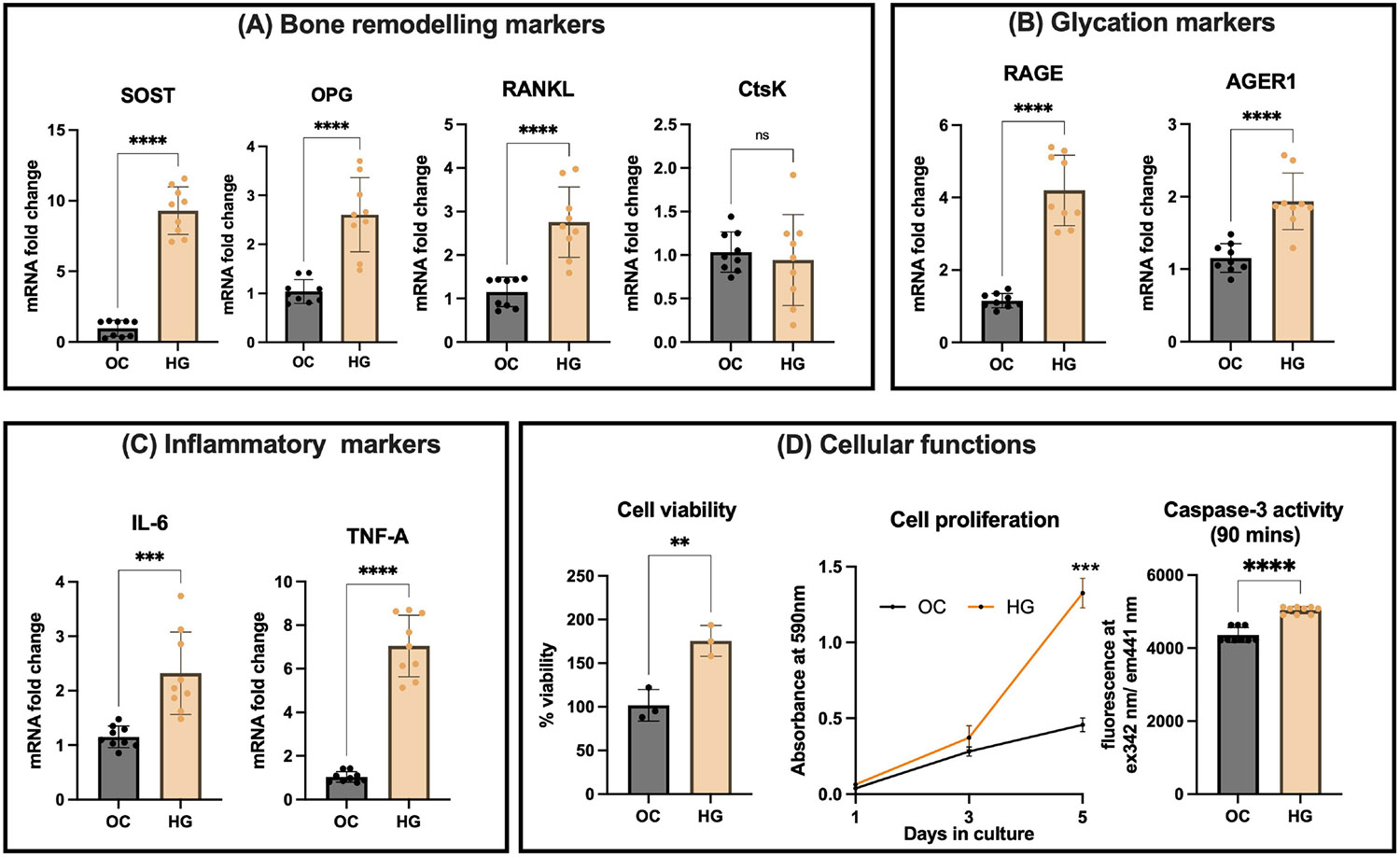
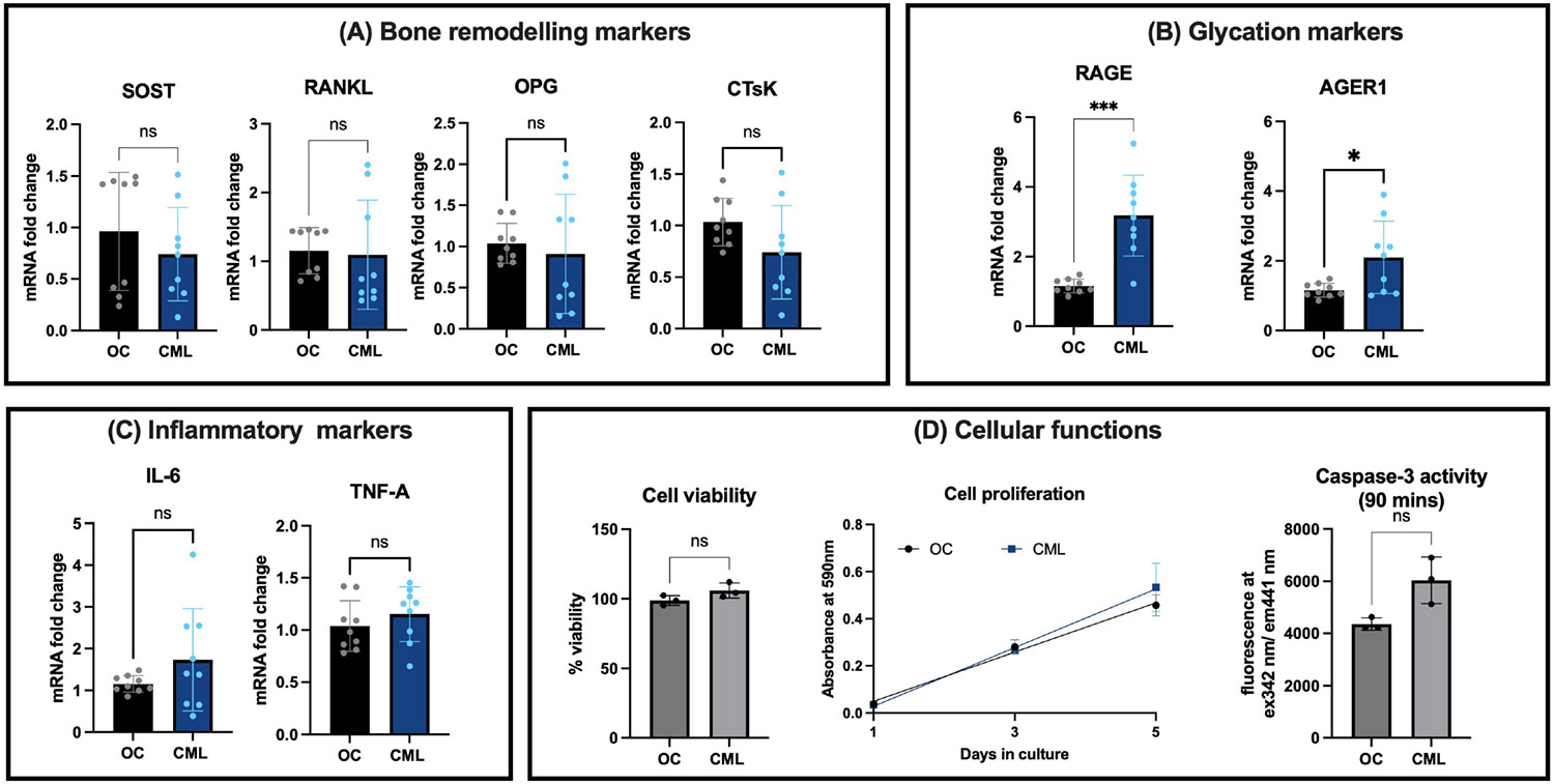
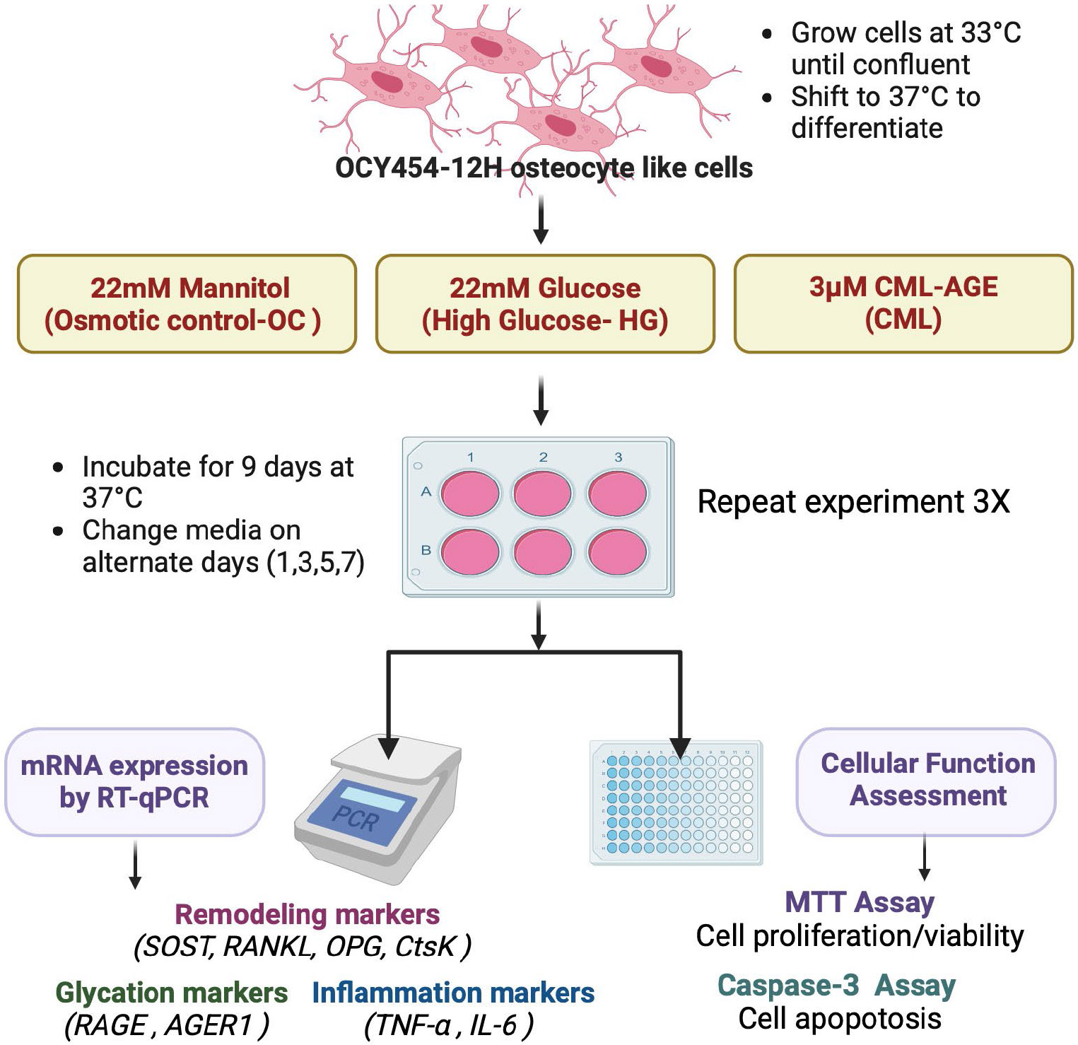
尽管骨密度正常或升高,但糖尿病(DM)与骨脆性增加有关,部分原因是骨组织中晚期糖基化终产物(AGEs)的积累。AGEs,如羧甲基赖氨酸(CML),通过激活晚期糖基化终产物(RAGE)受体,引发氧化应激和炎症反应,损害骨细胞功能。本研究旨在探讨高糖(HG)和CML对骨重塑、糖基化、炎症标志物和骨细胞功能的影响。以小鼠骨细胞系OCY454-12H为实验对象,分别用HG (30 mM葡萄糖)或3 μM CML处理细胞,模拟糖尿病状态。我们使用定量PCR和功能分析评估骨重塑标志物(SOST、RANKL、OPG、CTsK)、糖基化标志物(RAGE、AGER1)、炎症因子(IL-6、TNF-α)和细胞功能(包括增殖、活力和凋亡)的表达。HG处理导致SOST表达增加10倍(9.3比0.9,p≤0.0001),RANKL表达增加2.4倍(2.75比1.15,p≤0.0001),OPG同时增加2倍(2.60比1.04,p≤0.0001)。RANKL/OPG比值保持不变(p = 0.15)。HG还显著增加RAGE表达3.67倍(4.20 vs. 1.15, p≤0.0001),AGER1表达1.65倍(1.94 vs. 1.15, p≤0.0001),IL-6表达增加2.02倍(2.32 vs. 1.12, p≤0.001),TNF-α表达增加7.35倍(7.04 vs. 1.04, p≤0.0001)。HG处理下细胞活力和增殖显著提高,同时caspase-3活性升高,表明细胞凋亡增强。相比之下,CML暴露显著上调RAGE (3.18 vs. 1.15, p≤0.0001)和AGER1 (2.10 vs. 1.14, p = 0.028),但在生理浓度下对骨重塑标志物、炎症细胞因子或细胞功能没有显著影响。我们的研究结果表明,HG通过改变骨重塑、糖基化和炎症途径来破坏骨细胞功能,而CML在生理水平上选择性地激活糖基化标记物,而不会引起更广泛的细胞功能障碍。这些结果强调了AGE-RAGE轴在糖尿病骨脆性中的作用,并强调了未来体内研究探索针对骨中AGE积累和RAGE信号的治疗策略的必要性。
Effect of High Glucose and Carboxymethyl-Lysine on Osteocyte Gene Expression.
Diabetes mellitus (DM) is associated with increased bone fragility despite normal or elevated bone mineral density, partially due to the accumulation of advanced glycation end products (AGEs) in bone tissue. AGEs, such as carboxymethyl lysine (CML), impair osteocyte function by activating the receptor for advanced glycation end products (RAGE), triggering oxidative stress and inflammatory responses. This study aimed to investigate the effects of high glucose (HG) and CML on bone remodeling, glycation, inflammatory markers, and cellular functions in osteocytes. Using the murine osteocyte cell line OCY454-12H, we treated cells with HG (30 mM glucose) or 3 μM CML to simulate diabetic conditions. We assessed the expression of bone remodeling markers (SOST, RANKL, OPG, CTsK), glycation markers (RAGE, AGER1), inflammatory cytokines (IL-6, TNF-α), and cellular functions, including proliferation, viability, and apoptosis, using quantitative PCR and functional assays. HG treatment resulted in a 10-fold increase in SOST expression (9.3 vs. 0.9, p ≤ 0.0001) and a 2.4-fold increase in RANKL expression (2.75 vs. 1.15, p ≤ 0.0001), with a concurrent 2-fold increase in OPG (2.60 vs. 1.04, p ≤ 0.0001). The RANKL/OPG ratio remained unchanged (p = 0.15). HG also significantly increased RAGE expression by 3.67-fold (4.20 vs. 1.15, p ≤ 0.0001) and AGER1 by 1.65-fold (1.94 vs. 1.15, p ≤ 0.0001), along with a 2.02-fold increase in IL-6 (2.32 vs. 1.12, p ≤ 0.001) and a 7.35-fold increase in TNF-α (7.04 vs. 1.04, p ≤ 0.0001). Cell viability and proliferation were significantly higher under HG, accompanied by increased caspase-3 activity, indicating enhanced apoptosis. In contrast, CML exposure significantly upregulated RAGE (3.18 vs. 1.15, p ≤ 0.0001) and AGER1 (2.10 vs. 1.14, p = 0.028) but had no significant effects on bone remodeling markers, inflammatory cytokines, or cellular functions at physiological concentrations. Our findings demonstrate that HG disrupts osteocyte function by altering bone remodeling, glycation, and inflammatory pathways, while CML at physiological levels selectively activates glycation markers without inducing broader cellular dysfunction. These results underscore the role of the AGE-RAGE axis in diabetic bone fragility and highlight the need for future in vivo studies to explore therapeutic strategies targeting AGE accumulation and RAGE signaling in bone.
 求助内容:
求助内容: 应助结果提醒方式:


