{"title":"高清晰度和自身荧光支气管镜成像评估鳞状细胞肺癌新辅助免疫化疗后上皮细胞的变化:1例报告。","authors":"Kei Morikawa, Koji Kojima, Hideki Marushima, Yoshiya Sugiura, Junki Koike, Hisashi Saji, Masamichi Mineshita","doi":"10.1111/1759-7714.70096","DOIUrl":null,"url":null,"abstract":"<p><p>In recent years, perioperative immune checkpoint inhibitors have become indicated for early-stage lung cancer, emphasizing the importance of high-resolution endoscopic evaluation of preoperative drug therapy. At the initial evaluation, a male patient in his 60s presented with a primary lesion obstructing the right upper lobe bronchus. After three courses of neoadjuvant immunochemotherapy, chest computed tomography and endoscopic examinations showed a near-complete response. Narrow-band imaging indicated that subepithelial vascular regularity and distribution patterns were within normal limits. However, autofluorescence imaging (AFI) revealed a magenta-colored area on the bronchial epithelium corresponding to the initial lesion site. Two months later, the magenta coloration faded, suggesting pathological normalization of the bronchial epithelium thickening. AFI enabled visualization of tumor progression in the bronchi otherwise completely obstructed by the lesion, potentially offering valuable information to determine bronchial resection lines during surgery.</p>","PeriodicalId":23338,"journal":{"name":"Thoracic Cancer","volume":"16 10","pages":"e70096"},"PeriodicalIF":2.3000,"publicationDate":"2025-05-01","publicationTypes":"Journal Article","fieldsOfStudy":null,"isOpenAccess":false,"openAccessPdf":"https://www.ncbi.nlm.nih.gov/pmc/articles/PMC12095845/pdf/","citationCount":"0","resultStr":"{\"title\":\"High-Definition and Autofluorescence Bronchoscopic Imaging for Evaluating Epithelial Changes in Squamous Cell Lung Cancer After Neoadjuvant Immunochemotherapy: A Case Report.\",\"authors\":\"Kei Morikawa, Koji Kojima, Hideki Marushima, Yoshiya Sugiura, Junki Koike, Hisashi Saji, Masamichi Mineshita\",\"doi\":\"10.1111/1759-7714.70096\",\"DOIUrl\":null,\"url\":null,\"abstract\":\"<p><p>In recent years, perioperative immune checkpoint inhibitors have become indicated for early-stage lung cancer, emphasizing the importance of high-resolution endoscopic evaluation of preoperative drug therapy. At the initial evaluation, a male patient in his 60s presented with a primary lesion obstructing the right upper lobe bronchus. After three courses of neoadjuvant immunochemotherapy, chest computed tomography and endoscopic examinations showed a near-complete response. Narrow-band imaging indicated that subepithelial vascular regularity and distribution patterns were within normal limits. However, autofluorescence imaging (AFI) revealed a magenta-colored area on the bronchial epithelium corresponding to the initial lesion site. Two months later, the magenta coloration faded, suggesting pathological normalization of the bronchial epithelium thickening. AFI enabled visualization of tumor progression in the bronchi otherwise completely obstructed by the lesion, potentially offering valuable information to determine bronchial resection lines during surgery.</p>\",\"PeriodicalId\":23338,\"journal\":{\"name\":\"Thoracic Cancer\",\"volume\":\"16 10\",\"pages\":\"e70096\"},\"PeriodicalIF\":2.3000,\"publicationDate\":\"2025-05-01\",\"publicationTypes\":\"Journal Article\",\"fieldsOfStudy\":null,\"isOpenAccess\":false,\"openAccessPdf\":\"https://www.ncbi.nlm.nih.gov/pmc/articles/PMC12095845/pdf/\",\"citationCount\":\"0\",\"resultStr\":null,\"platform\":\"Semanticscholar\",\"paperid\":null,\"PeriodicalName\":\"Thoracic Cancer\",\"FirstCategoryId\":\"3\",\"ListUrlMain\":\"https://doi.org/10.1111/1759-7714.70096\",\"RegionNum\":3,\"RegionCategory\":\"医学\",\"ArticlePicture\":[],\"TitleCN\":null,\"AbstractTextCN\":null,\"PMCID\":null,\"EPubDate\":\"\",\"PubModel\":\"\",\"JCR\":\"Q3\",\"JCRName\":\"ONCOLOGY\",\"Score\":null,\"Total\":0}","platform":"Semanticscholar","paperid":null,"PeriodicalName":"Thoracic Cancer","FirstCategoryId":"3","ListUrlMain":"https://doi.org/10.1111/1759-7714.70096","RegionNum":3,"RegionCategory":"医学","ArticlePicture":[],"TitleCN":null,"AbstractTextCN":null,"PMCID":null,"EPubDate":"","PubModel":"","JCR":"Q3","JCRName":"ONCOLOGY","Score":null,"Total":0}
High-Definition and Autofluorescence Bronchoscopic Imaging for Evaluating Epithelial Changes in Squamous Cell Lung Cancer After Neoadjuvant Immunochemotherapy: A Case Report.
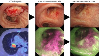
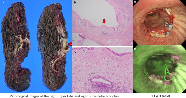
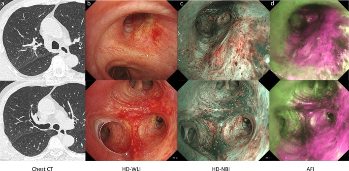
In recent years, perioperative immune checkpoint inhibitors have become indicated for early-stage lung cancer, emphasizing the importance of high-resolution endoscopic evaluation of preoperative drug therapy. At the initial evaluation, a male patient in his 60s presented with a primary lesion obstructing the right upper lobe bronchus. After three courses of neoadjuvant immunochemotherapy, chest computed tomography and endoscopic examinations showed a near-complete response. Narrow-band imaging indicated that subepithelial vascular regularity and distribution patterns were within normal limits. However, autofluorescence imaging (AFI) revealed a magenta-colored area on the bronchial epithelium corresponding to the initial lesion site. Two months later, the magenta coloration faded, suggesting pathological normalization of the bronchial epithelium thickening. AFI enabled visualization of tumor progression in the bronchi otherwise completely obstructed by the lesion, potentially offering valuable information to determine bronchial resection lines during surgery.
期刊介绍:
Thoracic Cancer aims to facilitate international collaboration and exchange of comprehensive and cutting-edge information on basic, translational, and applied clinical research in lung cancer, esophageal cancer, mediastinal cancer, breast cancer and other thoracic malignancies. Prevention, treatment and research relevant to Asia-Pacific is a focus area, but submissions from all regions are welcomed. The editors encourage contributions relevant to prevention, general thoracic surgery, medical oncology, radiology, radiation medicine, pathology, basic cancer research, as well as epidemiological and translational studies in thoracic cancer. Thoracic Cancer is the official publication of the Chinese Society of Lung Cancer, International Chinese Society of Thoracic Surgery and is endorsed by the Korean Association for the Study of Lung Cancer and the Hong Kong Cancer Therapy Society.
The Journal publishes a range of article types including: Editorials, Invited Reviews, Mini Reviews, Original Articles, Clinical Guidelines, Technological Notes, Imaging in thoracic cancer, Meeting Reports, Case Reports, Letters to the Editor, Commentaries, and Brief Reports.
 求助内容:
求助内容: 应助结果提醒方式:


