Sara Alrumaih, Nouf Alshibani, Rita Khounganian, Fahad A Alshehri, Eman Allam, Reem Alkattan
{"title":"解决蛋白E1与大鼠颅骨缺损的综合组织学分析。","authors":"Sara Alrumaih, Nouf Alshibani, Rita Khounganian, Fahad A Alshehri, Eman Allam, Reem Alkattan","doi":"10.1007/s44445-025-00003-4","DOIUrl":null,"url":null,"abstract":"<p><p>Bone loss, linked with numerous oral conditions such as periodontal diseases and periimplantitis, poses a significant challenge for dental clinicians. The current study evaluated the in vivo effects of local application of Resolvin E1 (RvE1) on bone regeneration in critical size calvarial defects in rats. Thirty female Wistar rats with 5 mm induced calvarial defects were randomly allocated into four groups: no treatment (negative control, n = 5), treatment using bovine bone grafts (positive control, n = 5), treatment using local delivery of RvE1 (n = 11) and treatment using RvE1 mixed with bovine bone graft (n = 9). After 12 weeks, the animals were sacrificed and the calvarial defects with the adjacent tissues were sectioned en-bloc and prepared for histological examination. A comprehensive qualitative and quantitative histological examination was performed to assess bone regeneration and its relation to the defect, the presence of remnant bone and RvE1 particles, the integration of the native bone with the newly formed bone, bone density and bony trabeculae, the inflammatory reaction, the connective tissue bridging in the defect, and the encapsulating fibrous tissue. Signs of neovascularization, increased cellularity, lack of the organized lamellated appearance of mature bone, disorganized arrangement of osteocytes, osteoblasts and osteoclasts were also assessed. Comparisons of the quantitative values between all studied groups indicated statistically significant differences (p ≤ 0.05) in all parameters except for the increased cellularity and granulation tissue. Histological findings indicate that RvE1 with adjunct bone graft significantly enhanced the bone formation compared to RvE1 or bovine graft alone.</p>","PeriodicalId":47246,"journal":{"name":"Saudi Dental Journal","volume":"37 1-3","pages":"2"},"PeriodicalIF":2.3000,"publicationDate":"2025-04-14","publicationTypes":"Journal Article","fieldsOfStudy":null,"isOpenAccess":false,"openAccessPdf":"https://www.ncbi.nlm.nih.gov/pmc/articles/PMC12133619/pdf/","citationCount":"0","resultStr":"{\"title\":\"Resolvin E1 and calvarial defects in rats: a comprehensive histological analysis.\",\"authors\":\"Sara Alrumaih, Nouf Alshibani, Rita Khounganian, Fahad A Alshehri, Eman Allam, Reem Alkattan\",\"doi\":\"10.1007/s44445-025-00003-4\",\"DOIUrl\":null,\"url\":null,\"abstract\":\"<p><p>Bone loss, linked with numerous oral conditions such as periodontal diseases and periimplantitis, poses a significant challenge for dental clinicians. The current study evaluated the in vivo effects of local application of Resolvin E1 (RvE1) on bone regeneration in critical size calvarial defects in rats. Thirty female Wistar rats with 5 mm induced calvarial defects were randomly allocated into four groups: no treatment (negative control, n = 5), treatment using bovine bone grafts (positive control, n = 5), treatment using local delivery of RvE1 (n = 11) and treatment using RvE1 mixed with bovine bone graft (n = 9). After 12 weeks, the animals were sacrificed and the calvarial defects with the adjacent tissues were sectioned en-bloc and prepared for histological examination. A comprehensive qualitative and quantitative histological examination was performed to assess bone regeneration and its relation to the defect, the presence of remnant bone and RvE1 particles, the integration of the native bone with the newly formed bone, bone density and bony trabeculae, the inflammatory reaction, the connective tissue bridging in the defect, and the encapsulating fibrous tissue. Signs of neovascularization, increased cellularity, lack of the organized lamellated appearance of mature bone, disorganized arrangement of osteocytes, osteoblasts and osteoclasts were also assessed. Comparisons of the quantitative values between all studied groups indicated statistically significant differences (p ≤ 0.05) in all parameters except for the increased cellularity and granulation tissue. Histological findings indicate that RvE1 with adjunct bone graft significantly enhanced the bone formation compared to RvE1 or bovine graft alone.</p>\",\"PeriodicalId\":47246,\"journal\":{\"name\":\"Saudi Dental Journal\",\"volume\":\"37 1-3\",\"pages\":\"2\"},\"PeriodicalIF\":2.3000,\"publicationDate\":\"2025-04-14\",\"publicationTypes\":\"Journal Article\",\"fieldsOfStudy\":null,\"isOpenAccess\":false,\"openAccessPdf\":\"https://www.ncbi.nlm.nih.gov/pmc/articles/PMC12133619/pdf/\",\"citationCount\":\"0\",\"resultStr\":null,\"platform\":\"Semanticscholar\",\"paperid\":null,\"PeriodicalName\":\"Saudi Dental Journal\",\"FirstCategoryId\":\"1085\",\"ListUrlMain\":\"https://doi.org/10.1007/s44445-025-00003-4\",\"RegionNum\":0,\"RegionCategory\":null,\"ArticlePicture\":[],\"TitleCN\":null,\"AbstractTextCN\":null,\"PMCID\":null,\"EPubDate\":\"\",\"PubModel\":\"\",\"JCR\":\"Q3\",\"JCRName\":\"DENTISTRY, ORAL SURGERY & MEDICINE\",\"Score\":null,\"Total\":0}","platform":"Semanticscholar","paperid":null,"PeriodicalName":"Saudi Dental Journal","FirstCategoryId":"1085","ListUrlMain":"https://doi.org/10.1007/s44445-025-00003-4","RegionNum":0,"RegionCategory":null,"ArticlePicture":[],"TitleCN":null,"AbstractTextCN":null,"PMCID":null,"EPubDate":"","PubModel":"","JCR":"Q3","JCRName":"DENTISTRY, ORAL SURGERY & MEDICINE","Score":null,"Total":0}
Resolvin E1 and calvarial defects in rats: a comprehensive histological analysis.
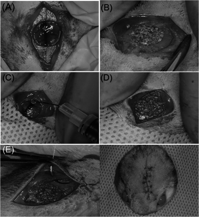
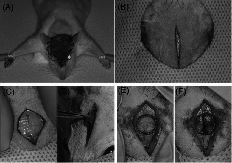
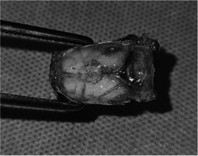
Bone loss, linked with numerous oral conditions such as periodontal diseases and periimplantitis, poses a significant challenge for dental clinicians. The current study evaluated the in vivo effects of local application of Resolvin E1 (RvE1) on bone regeneration in critical size calvarial defects in rats. Thirty female Wistar rats with 5 mm induced calvarial defects were randomly allocated into four groups: no treatment (negative control, n = 5), treatment using bovine bone grafts (positive control, n = 5), treatment using local delivery of RvE1 (n = 11) and treatment using RvE1 mixed with bovine bone graft (n = 9). After 12 weeks, the animals were sacrificed and the calvarial defects with the adjacent tissues were sectioned en-bloc and prepared for histological examination. A comprehensive qualitative and quantitative histological examination was performed to assess bone regeneration and its relation to the defect, the presence of remnant bone and RvE1 particles, the integration of the native bone with the newly formed bone, bone density and bony trabeculae, the inflammatory reaction, the connective tissue bridging in the defect, and the encapsulating fibrous tissue. Signs of neovascularization, increased cellularity, lack of the organized lamellated appearance of mature bone, disorganized arrangement of osteocytes, osteoblasts and osteoclasts were also assessed. Comparisons of the quantitative values between all studied groups indicated statistically significant differences (p ≤ 0.05) in all parameters except for the increased cellularity and granulation tissue. Histological findings indicate that RvE1 with adjunct bone graft significantly enhanced the bone formation compared to RvE1 or bovine graft alone.
期刊介绍:
Saudi Dental Journal is an English language, peer-reviewed scholarly publication in the area of dentistry. Saudi Dental Journal publishes original research and reviews on, but not limited to: • dental disease • clinical trials • dental equipment • new and experimental techniques • epidemiology and oral health • restorative dentistry • periodontology • endodontology • prosthodontics • paediatric dentistry • orthodontics and dental education Saudi Dental Journal is the official publication of the Saudi Dental Society and is published by King Saud University in collaboration with Elsevier and is edited by an international group of eminent researchers.
 求助内容:
求助内容: 应助结果提醒方式:


