Yan Zhang, Lu Liang, Huachong Ma, Jiagang Han, Xiuzhang Lv, Huiyu Ge
{"title":"经直肠双平面超声测量直肠肿瘤最低边界至肛门边缘距离的扩展视场成像评价。","authors":"Yan Zhang, Lu Liang, Huachong Ma, Jiagang Han, Xiuzhang Lv, Huiyu Ge","doi":"10.1055/a-2569-6939","DOIUrl":null,"url":null,"abstract":"<p><strong>Purpose: </strong>This study aimed to measure the precise distance from the lowest boundary of a rectal tumor to the anal verge (DTAV) in patients with rectal cancer.</p><p><strong>Materials and methods: </strong>A retrospective analysis was performed on clinical data from 70 rectal cancer patients. DTAV measurements were collected using transrectal biplane ultrasound, MRI, and colonoscopy.</p><p><strong>Results: </strong>The difference in DTAV measurements between the mean DTAV value obtained by ultrasound (US <sub>mean</sub> ) and colonoscopy exhibited a difference of 0.22 cm. In contrast, the difference between US <sub>mean</sub> and MRI was 0.48 cm, while the difference between MRI and colonoscopy was -0.26 cm. The ICC for DTAV measurements demonstrated excellent agreement, with values of 0.948 between US <sub>mean</sub> and MRI, 0.942 between US <sub>mean</sub> and colonoscopy, and 0.943 between MRI and colonoscopy. The minimum DTAV value obtained by ultrasound (US <sub>min</sub> ) was 5.05 cm, the middle DTAV value obtained by ultrasound (US <sub>mid</sub> ) was 5.10 cm, and the maximum DTAV value obtained by ultrasound (US <sub>max</sub> ) was 5.30 cm. Notably, the median values of the differences in DTAV measurements between US <sub>max</sub> and US <sub>min</sub> , US <sub>max</sub> and US <sub>mid</sub> , as well as US <sub>mid</sub> and US <sub>min</sub> , were 0.2 cm, 0.1 cm, and 0.1 cm, respectively. Furthermore, the consistency of DTAV measurements between US <sub>min</sub> and US <sub>mid</sub> , US <sub>max</sub> and US <sub>mid</sub> , as well as US <sub>min</sub> and US <sub>max</sub> was excellent, with all ICC values reaching 0.999. Additionally, the radiologist's reassessment of MRI DTAV data showed excellent consistency with the original results, with an ICC value of 0.985.</p><p><strong>Conclusion: </strong>Transrectal biplane ultrasound utilizing EFOV imaging technology exhibited both accuracy and reproducibility for measuring DTAV. This approach provided a highly efficient and practical clinical tool for DTAV measurement.</p>","PeriodicalId":44852,"journal":{"name":"Ultrasound International Open","volume":"11 ","pages":"a25696939"},"PeriodicalIF":1.6000,"publicationDate":"2025-05-05","publicationTypes":"Journal Article","fieldsOfStudy":null,"isOpenAccess":false,"openAccessPdf":"https://www.ncbi.nlm.nih.gov/pmc/articles/PMC12090980/pdf/","citationCount":"0","resultStr":"{\"title\":\"Evaluating Extended Field of View Imaging for Measuring Rectal Tumor Lowest Boundary to Anal Verge Distance via Transrectal Biplane Ultrasound.\",\"authors\":\"Yan Zhang, Lu Liang, Huachong Ma, Jiagang Han, Xiuzhang Lv, Huiyu Ge\",\"doi\":\"10.1055/a-2569-6939\",\"DOIUrl\":null,\"url\":null,\"abstract\":\"<p><strong>Purpose: </strong>This study aimed to measure the precise distance from the lowest boundary of a rectal tumor to the anal verge (DTAV) in patients with rectal cancer.</p><p><strong>Materials and methods: </strong>A retrospective analysis was performed on clinical data from 70 rectal cancer patients. DTAV measurements were collected using transrectal biplane ultrasound, MRI, and colonoscopy.</p><p><strong>Results: </strong>The difference in DTAV measurements between the mean DTAV value obtained by ultrasound (US <sub>mean</sub> ) and colonoscopy exhibited a difference of 0.22 cm. In contrast, the difference between US <sub>mean</sub> and MRI was 0.48 cm, while the difference between MRI and colonoscopy was -0.26 cm. The ICC for DTAV measurements demonstrated excellent agreement, with values of 0.948 between US <sub>mean</sub> and MRI, 0.942 between US <sub>mean</sub> and colonoscopy, and 0.943 between MRI and colonoscopy. The minimum DTAV value obtained by ultrasound (US <sub>min</sub> ) was 5.05 cm, the middle DTAV value obtained by ultrasound (US <sub>mid</sub> ) was 5.10 cm, and the maximum DTAV value obtained by ultrasound (US <sub>max</sub> ) was 5.30 cm. Notably, the median values of the differences in DTAV measurements between US <sub>max</sub> and US <sub>min</sub> , US <sub>max</sub> and US <sub>mid</sub> , as well as US <sub>mid</sub> and US <sub>min</sub> , were 0.2 cm, 0.1 cm, and 0.1 cm, respectively. Furthermore, the consistency of DTAV measurements between US <sub>min</sub> and US <sub>mid</sub> , US <sub>max</sub> and US <sub>mid</sub> , as well as US <sub>min</sub> and US <sub>max</sub> was excellent, with all ICC values reaching 0.999. Additionally, the radiologist's reassessment of MRI DTAV data showed excellent consistency with the original results, with an ICC value of 0.985.</p><p><strong>Conclusion: </strong>Transrectal biplane ultrasound utilizing EFOV imaging technology exhibited both accuracy and reproducibility for measuring DTAV. This approach provided a highly efficient and practical clinical tool for DTAV measurement.</p>\",\"PeriodicalId\":44852,\"journal\":{\"name\":\"Ultrasound International Open\",\"volume\":\"11 \",\"pages\":\"a25696939\"},\"PeriodicalIF\":1.6000,\"publicationDate\":\"2025-05-05\",\"publicationTypes\":\"Journal Article\",\"fieldsOfStudy\":null,\"isOpenAccess\":false,\"openAccessPdf\":\"https://www.ncbi.nlm.nih.gov/pmc/articles/PMC12090980/pdf/\",\"citationCount\":\"0\",\"resultStr\":null,\"platform\":\"Semanticscholar\",\"paperid\":null,\"PeriodicalName\":\"Ultrasound International Open\",\"FirstCategoryId\":\"1085\",\"ListUrlMain\":\"https://doi.org/10.1055/a-2569-6939\",\"RegionNum\":0,\"RegionCategory\":null,\"ArticlePicture\":[],\"TitleCN\":null,\"AbstractTextCN\":null,\"PMCID\":null,\"EPubDate\":\"2025/1/1 0:00:00\",\"PubModel\":\"eCollection\",\"JCR\":\"Q3\",\"JCRName\":\"RADIOLOGY, NUCLEAR MEDICINE & MEDICAL IMAGING\",\"Score\":null,\"Total\":0}","platform":"Semanticscholar","paperid":null,"PeriodicalName":"Ultrasound International Open","FirstCategoryId":"1085","ListUrlMain":"https://doi.org/10.1055/a-2569-6939","RegionNum":0,"RegionCategory":null,"ArticlePicture":[],"TitleCN":null,"AbstractTextCN":null,"PMCID":null,"EPubDate":"2025/1/1 0:00:00","PubModel":"eCollection","JCR":"Q3","JCRName":"RADIOLOGY, NUCLEAR MEDICINE & MEDICAL IMAGING","Score":null,"Total":0}
Evaluating Extended Field of View Imaging for Measuring Rectal Tumor Lowest Boundary to Anal Verge Distance via Transrectal Biplane Ultrasound.
Purpose: This study aimed to measure the precise distance from the lowest boundary of a rectal tumor to the anal verge (DTAV) in patients with rectal cancer.
Materials and methods: A retrospective analysis was performed on clinical data from 70 rectal cancer patients. DTAV measurements were collected using transrectal biplane ultrasound, MRI, and colonoscopy.
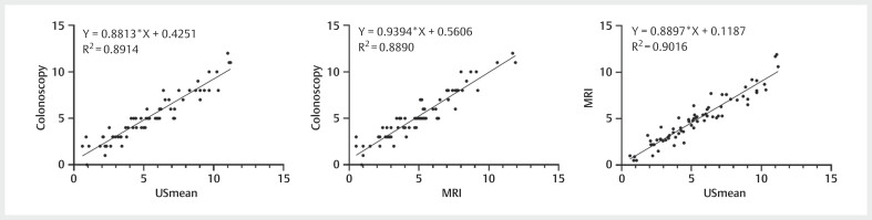
Results: The difference in DTAV measurements between the mean DTAV value obtained by ultrasound (US mean ) and colonoscopy exhibited a difference of 0.22 cm. In contrast, the difference between US mean and MRI was 0.48 cm, while the difference between MRI and colonoscopy was -0.26 cm. The ICC for DTAV measurements demonstrated excellent agreement, with values of 0.948 between US mean and MRI, 0.942 between US mean and colonoscopy, and 0.943 between MRI and colonoscopy. The minimum DTAV value obtained by ultrasound (US min ) was 5.05 cm, the middle DTAV value obtained by ultrasound (US mid ) was 5.10 cm, and the maximum DTAV value obtained by ultrasound (US max ) was 5.30 cm. Notably, the median values of the differences in DTAV measurements between US max and US min , US max and US mid , as well as US mid and US min , were 0.2 cm, 0.1 cm, and 0.1 cm, respectively. Furthermore, the consistency of DTAV measurements between US min and US mid , US max and US mid , as well as US min and US max was excellent, with all ICC values reaching 0.999. Additionally, the radiologist's reassessment of MRI DTAV data showed excellent consistency with the original results, with an ICC value of 0.985.
Conclusion: Transrectal biplane ultrasound utilizing EFOV imaging technology exhibited both accuracy and reproducibility for measuring DTAV. This approach provided a highly efficient and practical clinical tool for DTAV measurement.

 求助内容:
求助内容: 应助结果提醒方式:


