{"title":"经导管主动脉瓣植入术中自膨胀瓣膜的慢性扩张与瓣旁渗漏的关系。","authors":"Yuuki Muto, Daisuke Isomatsu, Yu Sato, Akihiko Sato, Takeshi Shimizu, Tomofumi Misaka, Masayoshi Oikawa, Atsushi Kobayashi, Akiomi Yoshihisa, Kazuhiko Nakazato, Takafumi Ishida, Hirofumi Sekino, Kenji Fukushima, Hiroshi Ito, Yasuchika Takeishi","doi":"10.1007/s12928-025-01140-7","DOIUrl":null,"url":null,"abstract":"<p><p>Paravalvular leakage (PVL) is a clinical concern in transcatheter aortic valve implantation (TAVI). Although PVL has been reported to gradually decrease after self-expandable valves (SEVs) implantation, the mechanism remains unclear. Furthermore, while gradual expansion of SEVs has been documented, studies assessing their morphological changes during the chronic phase are lacking. This study aims to evaluate whether SEVs expansion occurs during follow-up and if this expansion correlates with a reduction in PVL. We included 60 patients who underwent TAVI using SEVs (Evolut PRO/PRO+/FX) from October 2020 to August 2024. Morphological findings of SEVs were assessed using computed tomography (CT), and PVL was evaluated at discharge and chronic phase post-TAVI. PVL was graded on a five-point scale via transthoracic echocardiography, and the area ratio, defined as the ratio of the basal area of the implanted SEVs to that of phantom valves measured by CT, served as the index of SEVs expansion. Chronic expansion of SEVs was observed in all cases, with the area ratio increasing from 0.59 at discharge to 0.64 at the follow-up (P < 0.05). Among the 60 patients, 44 exhibited mild or greater PVL, with 17 showing improvement at the chronic phase. The degree of valve expansion (area ratio at chronic phase/area ratio at discharge) was significantly greater in the PVL improvement group compared to the non-improvement group (1.11 vs. 1.06, P < 0.05). This study demonstrates that SEVs expand during the chronic phase after TAVI, which could contribute to the reduction of PVL.</p>","PeriodicalId":9439,"journal":{"name":"Cardiovascular Intervention and Therapeutics","volume":" ","pages":"669-678"},"PeriodicalIF":5.8000,"publicationDate":"2025-07-01","publicationTypes":"Journal Article","fieldsOfStudy":null,"isOpenAccess":false,"openAccessPdf":"https://www.ncbi.nlm.nih.gov/pmc/articles/PMC12167277/pdf/","citationCount":"0","resultStr":"{\"title\":\"The relationship between chronic expansion of self-expandable valves and paravalvular leakage in transcatheter aortic valve implantation.\",\"authors\":\"Yuuki Muto, Daisuke Isomatsu, Yu Sato, Akihiko Sato, Takeshi Shimizu, Tomofumi Misaka, Masayoshi Oikawa, Atsushi Kobayashi, Akiomi Yoshihisa, Kazuhiko Nakazato, Takafumi Ishida, Hirofumi Sekino, Kenji Fukushima, Hiroshi Ito, Yasuchika Takeishi\",\"doi\":\"10.1007/s12928-025-01140-7\",\"DOIUrl\":null,\"url\":null,\"abstract\":\"<p><p>Paravalvular leakage (PVL) is a clinical concern in transcatheter aortic valve implantation (TAVI). Although PVL has been reported to gradually decrease after self-expandable valves (SEVs) implantation, the mechanism remains unclear. Furthermore, while gradual expansion of SEVs has been documented, studies assessing their morphological changes during the chronic phase are lacking. This study aims to evaluate whether SEVs expansion occurs during follow-up and if this expansion correlates with a reduction in PVL. We included 60 patients who underwent TAVI using SEVs (Evolut PRO/PRO+/FX) from October 2020 to August 2024. Morphological findings of SEVs were assessed using computed tomography (CT), and PVL was evaluated at discharge and chronic phase post-TAVI. PVL was graded on a five-point scale via transthoracic echocardiography, and the area ratio, defined as the ratio of the basal area of the implanted SEVs to that of phantom valves measured by CT, served as the index of SEVs expansion. Chronic expansion of SEVs was observed in all cases, with the area ratio increasing from 0.59 at discharge to 0.64 at the follow-up (P < 0.05). Among the 60 patients, 44 exhibited mild or greater PVL, with 17 showing improvement at the chronic phase. The degree of valve expansion (area ratio at chronic phase/area ratio at discharge) was significantly greater in the PVL improvement group compared to the non-improvement group (1.11 vs. 1.06, P < 0.05). This study demonstrates that SEVs expand during the chronic phase after TAVI, which could contribute to the reduction of PVL.</p>\",\"PeriodicalId\":9439,\"journal\":{\"name\":\"Cardiovascular Intervention and Therapeutics\",\"volume\":\" \",\"pages\":\"669-678\"},\"PeriodicalIF\":5.8000,\"publicationDate\":\"2025-07-01\",\"publicationTypes\":\"Journal Article\",\"fieldsOfStudy\":null,\"isOpenAccess\":false,\"openAccessPdf\":\"https://www.ncbi.nlm.nih.gov/pmc/articles/PMC12167277/pdf/\",\"citationCount\":\"0\",\"resultStr\":null,\"platform\":\"Semanticscholar\",\"paperid\":null,\"PeriodicalName\":\"Cardiovascular Intervention and Therapeutics\",\"FirstCategoryId\":\"1085\",\"ListUrlMain\":\"https://doi.org/10.1007/s12928-025-01140-7\",\"RegionNum\":0,\"RegionCategory\":null,\"ArticlePicture\":[],\"TitleCN\":null,\"AbstractTextCN\":null,\"PMCID\":null,\"EPubDate\":\"2025/5/20 0:00:00\",\"PubModel\":\"Epub\",\"JCR\":\"Q2\",\"JCRName\":\"CARDIAC & CARDIOVASCULAR SYSTEMS\",\"Score\":null,\"Total\":0}","platform":"Semanticscholar","paperid":null,"PeriodicalName":"Cardiovascular Intervention and Therapeutics","FirstCategoryId":"1085","ListUrlMain":"https://doi.org/10.1007/s12928-025-01140-7","RegionNum":0,"RegionCategory":null,"ArticlePicture":[],"TitleCN":null,"AbstractTextCN":null,"PMCID":null,"EPubDate":"2025/5/20 0:00:00","PubModel":"Epub","JCR":"Q2","JCRName":"CARDIAC & CARDIOVASCULAR SYSTEMS","Score":null,"Total":0}
The relationship between chronic expansion of self-expandable valves and paravalvular leakage in transcatheter aortic valve implantation.
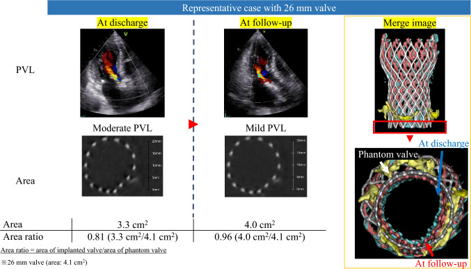
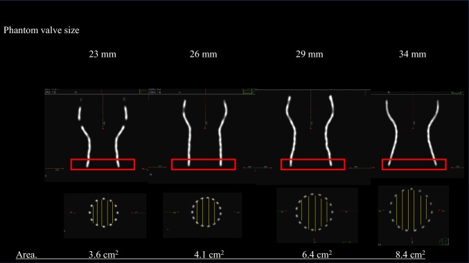
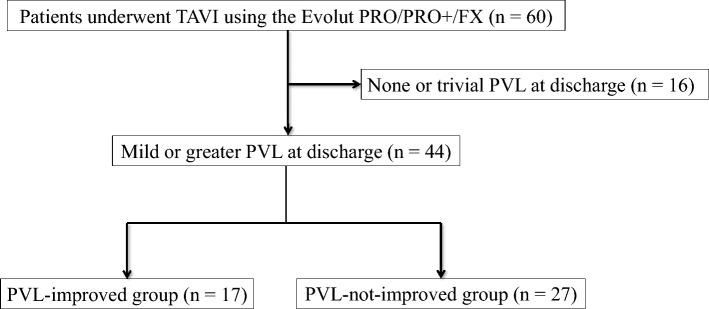
Paravalvular leakage (PVL) is a clinical concern in transcatheter aortic valve implantation (TAVI). Although PVL has been reported to gradually decrease after self-expandable valves (SEVs) implantation, the mechanism remains unclear. Furthermore, while gradual expansion of SEVs has been documented, studies assessing their morphological changes during the chronic phase are lacking. This study aims to evaluate whether SEVs expansion occurs during follow-up and if this expansion correlates with a reduction in PVL. We included 60 patients who underwent TAVI using SEVs (Evolut PRO/PRO+/FX) from October 2020 to August 2024. Morphological findings of SEVs were assessed using computed tomography (CT), and PVL was evaluated at discharge and chronic phase post-TAVI. PVL was graded on a five-point scale via transthoracic echocardiography, and the area ratio, defined as the ratio of the basal area of the implanted SEVs to that of phantom valves measured by CT, served as the index of SEVs expansion. Chronic expansion of SEVs was observed in all cases, with the area ratio increasing from 0.59 at discharge to 0.64 at the follow-up (P < 0.05). Among the 60 patients, 44 exhibited mild or greater PVL, with 17 showing improvement at the chronic phase. The degree of valve expansion (area ratio at chronic phase/area ratio at discharge) was significantly greater in the PVL improvement group compared to the non-improvement group (1.11 vs. 1.06, P < 0.05). This study demonstrates that SEVs expand during the chronic phase after TAVI, which could contribute to the reduction of PVL.
期刊介绍:
Cardiovascular Intervention and Therapeutics (CVIT) is an international journal covering the field of cardiovascular disease and includes cardiac (coronary and noncoronary) and peripheral interventions and therapeutics. Articles are subject to peer review and complete editorial evaluation prior to any decision regarding acceptability. CVIT is an official journal of The Japanese Association of Cardiovascular Intervention and Therapeutics.
 求助内容:
求助内容: 应助结果提醒方式:


