{"title":"磁共振成像评估黏液样脂肪肉瘤各成分与预后的相关性。","authors":"Jianjun Hua, Wenting Yang, Angcheng Li, Sisis Wang, Mingliang Ying","doi":"10.1177/02841851251337861","DOIUrl":null,"url":null,"abstract":"<p><p>BackgroundMyxoid liposarcoma (MLS) is a subtype of liposarcoma characterized by its myxoid stroma and adipocyte differentiation. MLS is prone to recurrence and metastasis. Magnetic resonance imaging (MRI) plays a crucial role in evaluating tumor characteristics, enabling accurate diagnosis, and predicting patient prognosis.PurposeTo analyze the components of MLS by MRI features and assess their correlation with prognosis.Material and MethodsA total of 20 patients with MLS who underwent MRI were retrospectively included. Tumor components were analyzed by MRI features, and their prognostic correlation was assessed. Patients were divided into good and poor prognosis groups based on postoperative follow-up.ResultsThe proportions of non-fatty/non-myxoid components in the good and poor prognosis groups were 15.00% (range = 10.00%-20.00%) and 70.00% (range = 52.50%-77.50%), respectively (<i>P</i> < 0.001). The proportion of myxoid composition also differed significantly between the two groups (75.00%, [range = 65.00%-85.00%] vs. 25.00% [range = 17.50%-42.50%]; <i>P</i> < 0.001). The good prognosis group had a greater mean apparent diffusion coefficient (ADC) value (1.66 ± 0.23 × 10<sup>-3</sup> mm<sup>2</sup>/s) and a lower mean ADC low signal ratio (5.00% [range = 0%-10.00%]) in the non-fatty/non-myxoid areas than the poor group (1.21 ± 0.41 × 10<sup>-3</sup> mm<sup>2</sup>/s; 20.00% [range = 11.00%-39.00%]; <i>P</i> <i>=</i> 0.006 and <i>P</i> <i>=</i> 0.001). The differences in the percentages of patients with a component ratio <25% and >50% in both the non-fatty/non-myxoid and myxoid groups were significant (<i>P</i> < 0.001 and <i>P</i> <i>=</i> 0.005).ConclusionImaging features were closely associated with the histological components of MLS. The use of MRI features for assessing MLS components has important implications for prognostic prediction.</p>","PeriodicalId":7143,"journal":{"name":"Acta radiologica","volume":" ","pages":"999-1007"},"PeriodicalIF":1.1000,"publicationDate":"2025-09-01","publicationTypes":"Journal Article","fieldsOfStudy":null,"isOpenAccess":false,"openAccessPdf":"https://www.ncbi.nlm.nih.gov/pmc/articles/PMC12411680/pdf/","citationCount":"0","resultStr":"{\"title\":\"Magnetic resonance imaging assessing the correlation of components and prognosis in myxoid liposarcoma.\",\"authors\":\"Jianjun Hua, Wenting Yang, Angcheng Li, Sisis Wang, Mingliang Ying\",\"doi\":\"10.1177/02841851251337861\",\"DOIUrl\":null,\"url\":null,\"abstract\":\"<p><p>BackgroundMyxoid liposarcoma (MLS) is a subtype of liposarcoma characterized by its myxoid stroma and adipocyte differentiation. MLS is prone to recurrence and metastasis. Magnetic resonance imaging (MRI) plays a crucial role in evaluating tumor characteristics, enabling accurate diagnosis, and predicting patient prognosis.PurposeTo analyze the components of MLS by MRI features and assess their correlation with prognosis.Material and MethodsA total of 20 patients with MLS who underwent MRI were retrospectively included. Tumor components were analyzed by MRI features, and their prognostic correlation was assessed. Patients were divided into good and poor prognosis groups based on postoperative follow-up.ResultsThe proportions of non-fatty/non-myxoid components in the good and poor prognosis groups were 15.00% (range = 10.00%-20.00%) and 70.00% (range = 52.50%-77.50%), respectively (<i>P</i> < 0.001). The proportion of myxoid composition also differed significantly between the two groups (75.00%, [range = 65.00%-85.00%] vs. 25.00% [range = 17.50%-42.50%]; <i>P</i> < 0.001). The good prognosis group had a greater mean apparent diffusion coefficient (ADC) value (1.66 ± 0.23 × 10<sup>-3</sup> mm<sup>2</sup>/s) and a lower mean ADC low signal ratio (5.00% [range = 0%-10.00%]) in the non-fatty/non-myxoid areas than the poor group (1.21 ± 0.41 × 10<sup>-3</sup> mm<sup>2</sup>/s; 20.00% [range = 11.00%-39.00%]; <i>P</i> <i>=</i> 0.006 and <i>P</i> <i>=</i> 0.001). The differences in the percentages of patients with a component ratio <25% and >50% in both the non-fatty/non-myxoid and myxoid groups were significant (<i>P</i> < 0.001 and <i>P</i> <i>=</i> 0.005).ConclusionImaging features were closely associated with the histological components of MLS. The use of MRI features for assessing MLS components has important implications for prognostic prediction.</p>\",\"PeriodicalId\":7143,\"journal\":{\"name\":\"Acta radiologica\",\"volume\":\" \",\"pages\":\"999-1007\"},\"PeriodicalIF\":1.1000,\"publicationDate\":\"2025-09-01\",\"publicationTypes\":\"Journal Article\",\"fieldsOfStudy\":null,\"isOpenAccess\":false,\"openAccessPdf\":\"https://www.ncbi.nlm.nih.gov/pmc/articles/PMC12411680/pdf/\",\"citationCount\":\"0\",\"resultStr\":null,\"platform\":\"Semanticscholar\",\"paperid\":null,\"PeriodicalName\":\"Acta radiologica\",\"FirstCategoryId\":\"3\",\"ListUrlMain\":\"https://doi.org/10.1177/02841851251337861\",\"RegionNum\":4,\"RegionCategory\":\"医学\",\"ArticlePicture\":[],\"TitleCN\":null,\"AbstractTextCN\":null,\"PMCID\":null,\"EPubDate\":\"2025/5/21 0:00:00\",\"PubModel\":\"Epub\",\"JCR\":\"Q3\",\"JCRName\":\"RADIOLOGY, NUCLEAR MEDICINE & MEDICAL IMAGING\",\"Score\":null,\"Total\":0}","platform":"Semanticscholar","paperid":null,"PeriodicalName":"Acta radiologica","FirstCategoryId":"3","ListUrlMain":"https://doi.org/10.1177/02841851251337861","RegionNum":4,"RegionCategory":"医学","ArticlePicture":[],"TitleCN":null,"AbstractTextCN":null,"PMCID":null,"EPubDate":"2025/5/21 0:00:00","PubModel":"Epub","JCR":"Q3","JCRName":"RADIOLOGY, NUCLEAR MEDICINE & MEDICAL IMAGING","Score":null,"Total":0}
Magnetic resonance imaging assessing the correlation of components and prognosis in myxoid liposarcoma.
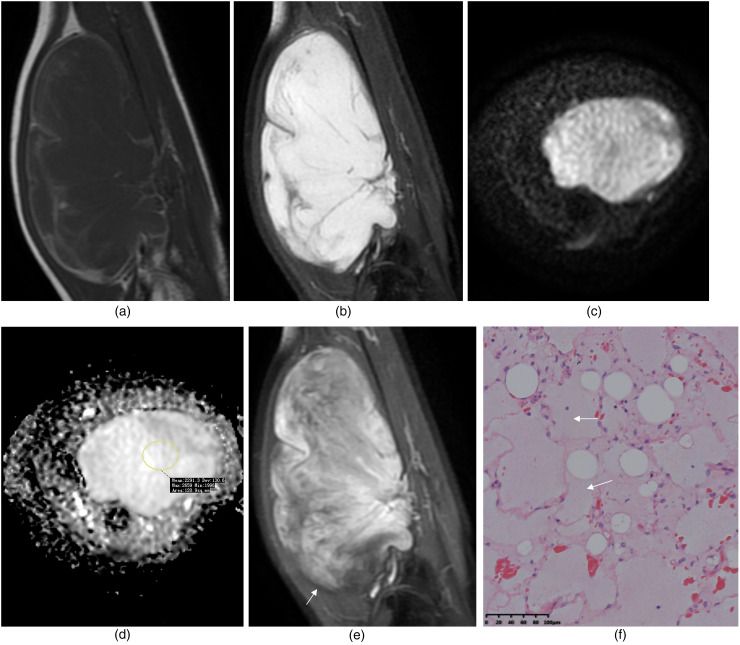
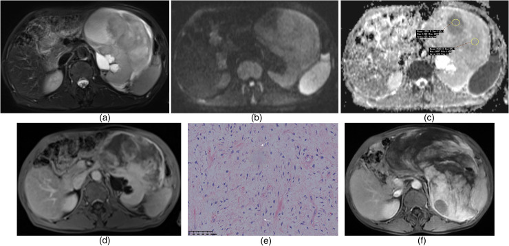
BackgroundMyxoid liposarcoma (MLS) is a subtype of liposarcoma characterized by its myxoid stroma and adipocyte differentiation. MLS is prone to recurrence and metastasis. Magnetic resonance imaging (MRI) plays a crucial role in evaluating tumor characteristics, enabling accurate diagnosis, and predicting patient prognosis.PurposeTo analyze the components of MLS by MRI features and assess their correlation with prognosis.Material and MethodsA total of 20 patients with MLS who underwent MRI were retrospectively included. Tumor components were analyzed by MRI features, and their prognostic correlation was assessed. Patients were divided into good and poor prognosis groups based on postoperative follow-up.ResultsThe proportions of non-fatty/non-myxoid components in the good and poor prognosis groups were 15.00% (range = 10.00%-20.00%) and 70.00% (range = 52.50%-77.50%), respectively (P < 0.001). The proportion of myxoid composition also differed significantly between the two groups (75.00%, [range = 65.00%-85.00%] vs. 25.00% [range = 17.50%-42.50%]; P < 0.001). The good prognosis group had a greater mean apparent diffusion coefficient (ADC) value (1.66 ± 0.23 × 10⁻³ mm2/s) and a lower mean ADC low signal ratio (5.00% [range = 0%-10.00%]) in the non-fatty/non-myxoid areas than the poor group (1.21 ± 0.41 × 10⁻³ mm2/s; 20.00% [range = 11.00%-39.00%]; P= 0.006 and P= 0.001). The differences in the percentages of patients with a component ratio <25% and >50% in both the non-fatty/non-myxoid and myxoid groups were significant (P < 0.001 and P= 0.005).ConclusionImaging features were closely associated with the histological components of MLS. The use of MRI features for assessing MLS components has important implications for prognostic prediction.
期刊介绍:
Acta Radiologica publishes articles on all aspects of radiology, from clinical radiology to experimental work. It is known for articles based on experimental work and contrast media research, giving priority to scientific original papers. The distinguished international editorial board also invite review articles, short communications and technical and instrumental notes.

 求助内容:
求助内容: 应助结果提醒方式:


