George Triantafyllou, Panagiotis Papadopoulos-Manolarakis, Panagiotis Papanagiotou, George Tsakotos, Maria Piagkou
{"title":"大脑中动脉的重复起源:一种罕见的变异。","authors":"George Triantafyllou, Panagiotis Papadopoulos-Manolarakis, Panagiotis Papanagiotou, George Tsakotos, Maria Piagkou","doi":"10.1007/s00276-025-03654-4","DOIUrl":null,"url":null,"abstract":"<p><p>The cerebral arterial circle exhibits considerable morphological variability. Variations in the middle cerebral artery (MCA) are infrequent occurrences that can be readily identified via imaging techniques. The current imaging report describes a rare variant of the MCA consisting of a duplicate origin, which was incidentally discovered in a 42-year-old female patient through computed tomography angiography. The duplicated origin of the MCA was identified on the left side of the cerebral arterial circle, forming an arterial ring with the temporopolar branch of the MCA originating from one of its limbs. The remainder of the arterial circle demonstrated no variants. It is essential to distinguish variants of the MCA without conflating them. The present variant is accurately characterized as a 'duplicate origin\", it should not be confused with fenestrations, and has a reported prevalence of 0.1%. Comprehending such arterial variations is vital prior to undertaking endovascular or neurosurgical procedures in the region.</p>","PeriodicalId":49461,"journal":{"name":"Surgical and Radiologic Anatomy","volume":"47 1","pages":"140"},"PeriodicalIF":1.2000,"publicationDate":"2025-05-19","publicationTypes":"Journal Article","fieldsOfStudy":null,"isOpenAccess":false,"openAccessPdf":"https://www.ncbi.nlm.nih.gov/pmc/articles/PMC12089186/pdf/","citationCount":"0","resultStr":"{\"title\":\"Duplicate origin of the middle cerebral artery: a rare variant.\",\"authors\":\"George Triantafyllou, Panagiotis Papadopoulos-Manolarakis, Panagiotis Papanagiotou, George Tsakotos, Maria Piagkou\",\"doi\":\"10.1007/s00276-025-03654-4\",\"DOIUrl\":null,\"url\":null,\"abstract\":\"<p><p>The cerebral arterial circle exhibits considerable morphological variability. Variations in the middle cerebral artery (MCA) are infrequent occurrences that can be readily identified via imaging techniques. The current imaging report describes a rare variant of the MCA consisting of a duplicate origin, which was incidentally discovered in a 42-year-old female patient through computed tomography angiography. The duplicated origin of the MCA was identified on the left side of the cerebral arterial circle, forming an arterial ring with the temporopolar branch of the MCA originating from one of its limbs. The remainder of the arterial circle demonstrated no variants. It is essential to distinguish variants of the MCA without conflating them. The present variant is accurately characterized as a 'duplicate origin\\\", it should not be confused with fenestrations, and has a reported prevalence of 0.1%. Comprehending such arterial variations is vital prior to undertaking endovascular or neurosurgical procedures in the region.</p>\",\"PeriodicalId\":49461,\"journal\":{\"name\":\"Surgical and Radiologic Anatomy\",\"volume\":\"47 1\",\"pages\":\"140\"},\"PeriodicalIF\":1.2000,\"publicationDate\":\"2025-05-19\",\"publicationTypes\":\"Journal Article\",\"fieldsOfStudy\":null,\"isOpenAccess\":false,\"openAccessPdf\":\"https://www.ncbi.nlm.nih.gov/pmc/articles/PMC12089186/pdf/\",\"citationCount\":\"0\",\"resultStr\":null,\"platform\":\"Semanticscholar\",\"paperid\":null,\"PeriodicalName\":\"Surgical and Radiologic Anatomy\",\"FirstCategoryId\":\"3\",\"ListUrlMain\":\"https://doi.org/10.1007/s00276-025-03654-4\",\"RegionNum\":4,\"RegionCategory\":\"医学\",\"ArticlePicture\":[],\"TitleCN\":null,\"AbstractTextCN\":null,\"PMCID\":null,\"EPubDate\":\"\",\"PubModel\":\"\",\"JCR\":\"Q2\",\"JCRName\":\"Medicine\",\"Score\":null,\"Total\":0}","platform":"Semanticscholar","paperid":null,"PeriodicalName":"Surgical and Radiologic Anatomy","FirstCategoryId":"3","ListUrlMain":"https://doi.org/10.1007/s00276-025-03654-4","RegionNum":4,"RegionCategory":"医学","ArticlePicture":[],"TitleCN":null,"AbstractTextCN":null,"PMCID":null,"EPubDate":"","PubModel":"","JCR":"Q2","JCRName":"Medicine","Score":null,"Total":0}
Duplicate origin of the middle cerebral artery: a rare variant.
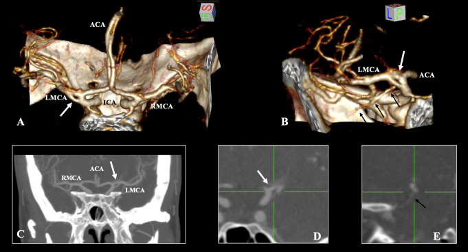
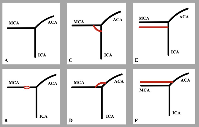
The cerebral arterial circle exhibits considerable morphological variability. Variations in the middle cerebral artery (MCA) are infrequent occurrences that can be readily identified via imaging techniques. The current imaging report describes a rare variant of the MCA consisting of a duplicate origin, which was incidentally discovered in a 42-year-old female patient through computed tomography angiography. The duplicated origin of the MCA was identified on the left side of the cerebral arterial circle, forming an arterial ring with the temporopolar branch of the MCA originating from one of its limbs. The remainder of the arterial circle demonstrated no variants. It is essential to distinguish variants of the MCA without conflating them. The present variant is accurately characterized as a 'duplicate origin", it should not be confused with fenestrations, and has a reported prevalence of 0.1%. Comprehending such arterial variations is vital prior to undertaking endovascular or neurosurgical procedures in the region.
期刊介绍:
Anatomy is a morphological science which cannot fail to interest the clinician. The practical application of anatomical research to clinical problems necessitates special adaptation and selectivity in choosing from numerous international works. Although there is a tendency to believe that meaningful advances in anatomy are unlikely, constant revision is necessary. Surgical and Radiologic Anatomy, the first international journal of Clinical anatomy has been created in this spirit.
Its goal is to serve clinicians, regardless of speciality-physicians, surgeons, radiologists or other specialists-as an indispensable aid with which they can improve their knowledge of anatomy. Each issue includes: Original papers, review articles, articles on the anatomical bases of medical, surgical and radiological techniques, articles of normal radiologic anatomy, brief reviews of anatomical publications of clinical interest.
Particular attention is given to high quality illustrations, which are indispensable for a better understanding of anatomical problems.
Surgical and Radiologic Anatomy is a journal written by anatomists for clinicians with a special interest in anatomy.

 求助内容:
求助内容: 应助结果提醒方式:


