Anna Luisa Silva Campos, Kezia de Souza Pinheiro, Matheus Leite Rassele, Marcos Rosa-Júnior
{"title":"神经隐球菌病的放射学表现谱:病例系列和系统回顾。","authors":"Anna Luisa Silva Campos, Kezia de Souza Pinheiro, Matheus Leite Rassele, Marcos Rosa-Júnior","doi":"10.1590/0100-3984.2024.0107","DOIUrl":null,"url":null,"abstract":"<p><p>This study involved a retrospective analysis of nine cases of neurocryptococcosis (eight from our institution and one from another institution) seen between May 2014 and May 2022, together with a systematic review of the literature indexed in the PubMed, Embase, and Lilacs databases. Clinical and radiological features of those cases were further refined via an additional comprehensive literature review. The following search string was employed: cryptococcosis AND central nervous system AND (magnetic resonance imaging OR X-ray computed tomography). The search was limited to articles published between July 1978 and May 2022. Two authors, working independently, searched for and selected studies that met the inclusion criteria, and another author reviewed conflicts in a blinded manner. We used Rayyan.ai software to organize the studies, and the review was structured in accordance with the 2020 Preferred Reporting Items for Systematic reviews and Meta-Analyses guidelines. Understanding the prevalence of different patterns of neurocryptococcosis is crucial for improving diagnosis and supporting decision-making in clinical practice. Our review of the literature demonstrated that imaging examinations are a valuable resource for early diagnosis, as well as for assessment of the initial extent and pattern of the disease.</p>","PeriodicalId":20842,"journal":{"name":"Radiologia Brasileira","volume":"58 ","pages":"e20240107"},"PeriodicalIF":0.0000,"publicationDate":"2025-05-01","publicationTypes":"Journal Article","fieldsOfStudy":null,"isOpenAccess":false,"openAccessPdf":"https://www.ncbi.nlm.nih.gov/pmc/articles/PMC12087350/pdf/","citationCount":"0","resultStr":"{\"title\":\"The spectrum of radiological findings in neurocryptococcosis: case series and systematic review.\",\"authors\":\"Anna Luisa Silva Campos, Kezia de Souza Pinheiro, Matheus Leite Rassele, Marcos Rosa-Júnior\",\"doi\":\"10.1590/0100-3984.2024.0107\",\"DOIUrl\":null,\"url\":null,\"abstract\":\"<p><p>This study involved a retrospective analysis of nine cases of neurocryptococcosis (eight from our institution and one from another institution) seen between May 2014 and May 2022, together with a systematic review of the literature indexed in the PubMed, Embase, and Lilacs databases. Clinical and radiological features of those cases were further refined via an additional comprehensive literature review. The following search string was employed: cryptococcosis AND central nervous system AND (magnetic resonance imaging OR X-ray computed tomography). The search was limited to articles published between July 1978 and May 2022. Two authors, working independently, searched for and selected studies that met the inclusion criteria, and another author reviewed conflicts in a blinded manner. We used Rayyan.ai software to organize the studies, and the review was structured in accordance with the 2020 Preferred Reporting Items for Systematic reviews and Meta-Analyses guidelines. Understanding the prevalence of different patterns of neurocryptococcosis is crucial for improving diagnosis and supporting decision-making in clinical practice. Our review of the literature demonstrated that imaging examinations are a valuable resource for early diagnosis, as well as for assessment of the initial extent and pattern of the disease.</p>\",\"PeriodicalId\":20842,\"journal\":{\"name\":\"Radiologia Brasileira\",\"volume\":\"58 \",\"pages\":\"e20240107\"},\"PeriodicalIF\":0.0000,\"publicationDate\":\"2025-05-01\",\"publicationTypes\":\"Journal Article\",\"fieldsOfStudy\":null,\"isOpenAccess\":false,\"openAccessPdf\":\"https://www.ncbi.nlm.nih.gov/pmc/articles/PMC12087350/pdf/\",\"citationCount\":\"0\",\"resultStr\":null,\"platform\":\"Semanticscholar\",\"paperid\":null,\"PeriodicalName\":\"Radiologia Brasileira\",\"FirstCategoryId\":\"1085\",\"ListUrlMain\":\"https://doi.org/10.1590/0100-3984.2024.0107\",\"RegionNum\":0,\"RegionCategory\":null,\"ArticlePicture\":[],\"TitleCN\":null,\"AbstractTextCN\":null,\"PMCID\":null,\"EPubDate\":\"2025/1/1 0:00:00\",\"PubModel\":\"eCollection\",\"JCR\":\"Q3\",\"JCRName\":\"Medicine\",\"Score\":null,\"Total\":0}","platform":"Semanticscholar","paperid":null,"PeriodicalName":"Radiologia Brasileira","FirstCategoryId":"1085","ListUrlMain":"https://doi.org/10.1590/0100-3984.2024.0107","RegionNum":0,"RegionCategory":null,"ArticlePicture":[],"TitleCN":null,"AbstractTextCN":null,"PMCID":null,"EPubDate":"2025/1/1 0:00:00","PubModel":"eCollection","JCR":"Q3","JCRName":"Medicine","Score":null,"Total":0}
The spectrum of radiological findings in neurocryptococcosis: case series and systematic review.
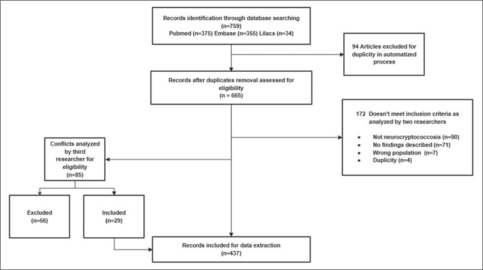
This study involved a retrospective analysis of nine cases of neurocryptococcosis (eight from our institution and one from another institution) seen between May 2014 and May 2022, together with a systematic review of the literature indexed in the PubMed, Embase, and Lilacs databases. Clinical and radiological features of those cases were further refined via an additional comprehensive literature review. The following search string was employed: cryptococcosis AND central nervous system AND (magnetic resonance imaging OR X-ray computed tomography). The search was limited to articles published between July 1978 and May 2022. Two authors, working independently, searched for and selected studies that met the inclusion criteria, and another author reviewed conflicts in a blinded manner. We used Rayyan.ai software to organize the studies, and the review was structured in accordance with the 2020 Preferred Reporting Items for Systematic reviews and Meta-Analyses guidelines. Understanding the prevalence of different patterns of neurocryptococcosis is crucial for improving diagnosis and supporting decision-making in clinical practice. Our review of the literature demonstrated that imaging examinations are a valuable resource for early diagnosis, as well as for assessment of the initial extent and pattern of the disease.

 求助内容:
求助内容: 应助结果提醒方式:


