Anouk Moens, Zoe Albersnagel, Marieke B Veenhof, Phebe N Adama van Scheltema, Esther Sikkel, Mariëtte J V Hoffer, Brigitte H W Faas, Dineke Westra, Ilse Feenstra, Emilia K Bijlsma, Gijs W E Santen, Corrie E Erasmus, Cacha M P C D Peeters-Scholte
{"title":"产前诊断胎儿脑室肿大进展的临床结局和危险因素:一项回顾性多中心研究。","authors":"Anouk Moens, Zoe Albersnagel, Marieke B Veenhof, Phebe N Adama van Scheltema, Esther Sikkel, Mariëtte J V Hoffer, Brigitte H W Faas, Dineke Westra, Ilse Feenstra, Emilia K Bijlsma, Gijs W E Santen, Corrie E Erasmus, Cacha M P C D Peeters-Scholte","doi":"10.1002/pd.6816","DOIUrl":null,"url":null,"abstract":"<p><strong>Objective: </strong>To investigate the clinical outcome of fetuses with ventriculomegaly (VM), and to identify risk factors for progression of fetal VM in order to improve prenatal counseling. This was a multicenter, retrospective cohort study, comprising 229 cases with VM.</p><p><strong>Methods: </strong>VM was classified as mild, moderate, or severe and isolated or non-isolated. Genetic data were collected. Differences between VM subgroups were described, and risk factors for progression of fetal VM were identified using logistic regression analysis. Outcome was defined as the percentage of live births, termination of pregnancy (TOP) and intra-uterine fetal demise (IUFD).</p><p><strong>Results: </strong>Of the 229 cases, 109 (47.6%) had mild VM, 60 (26.2%) moderate VM, and 60 (26.2%) severe VM. Progression of VM occurred in 45/153 cases (29.4%), half of which were in the group with severe VM. Dilatation of the 3rd ventricle and neural tube defects were risk factors for progression of VM. The percentage of live births (excluding cases with TOP and unknown outcome) was 93.1% (54/58) in mild VM, 78.6% (22/28) in moderate VM and 92.6% (25/27) in severe VM. In 12/229 cases (5.2%) IUFD occurred. Genetic analysis was performed in 143/229 (62.4%) of cases, showing (likely) pathogenic abnormalities in 41/143 (28.7%) cases, predominantly in mild, non-isolated VM.</p><p><strong>Conclusions: </strong>This study confirms the clinical relevance of additional genetic investigations in all types of fetal VMs. Further larger prospective research including clinical follow-up is needed to improve prenatal counseling.</p>","PeriodicalId":20387,"journal":{"name":"Prenatal Diagnosis","volume":" ","pages":"1089-1099"},"PeriodicalIF":2.7000,"publicationDate":"2025-08-01","publicationTypes":"Journal Article","fieldsOfStudy":null,"isOpenAccess":false,"openAccessPdf":"https://www.ncbi.nlm.nih.gov/pmc/articles/PMC12322251/pdf/","citationCount":"0","resultStr":"{\"title\":\"Clinical Outcome and Risk Factors for Progression of Prenatally Diagnosed Fetal Ventriculomegaly: A Retrospective Multicenter Study.\",\"authors\":\"Anouk Moens, Zoe Albersnagel, Marieke B Veenhof, Phebe N Adama van Scheltema, Esther Sikkel, Mariëtte J V Hoffer, Brigitte H W Faas, Dineke Westra, Ilse Feenstra, Emilia K Bijlsma, Gijs W E Santen, Corrie E Erasmus, Cacha M P C D Peeters-Scholte\",\"doi\":\"10.1002/pd.6816\",\"DOIUrl\":null,\"url\":null,\"abstract\":\"<p><strong>Objective: </strong>To investigate the clinical outcome of fetuses with ventriculomegaly (VM), and to identify risk factors for progression of fetal VM in order to improve prenatal counseling. This was a multicenter, retrospective cohort study, comprising 229 cases with VM.</p><p><strong>Methods: </strong>VM was classified as mild, moderate, or severe and isolated or non-isolated. Genetic data were collected. Differences between VM subgroups were described, and risk factors for progression of fetal VM were identified using logistic regression analysis. Outcome was defined as the percentage of live births, termination of pregnancy (TOP) and intra-uterine fetal demise (IUFD).</p><p><strong>Results: </strong>Of the 229 cases, 109 (47.6%) had mild VM, 60 (26.2%) moderate VM, and 60 (26.2%) severe VM. Progression of VM occurred in 45/153 cases (29.4%), half of which were in the group with severe VM. Dilatation of the 3rd ventricle and neural tube defects were risk factors for progression of VM. The percentage of live births (excluding cases with TOP and unknown outcome) was 93.1% (54/58) in mild VM, 78.6% (22/28) in moderate VM and 92.6% (25/27) in severe VM. In 12/229 cases (5.2%) IUFD occurred. Genetic analysis was performed in 143/229 (62.4%) of cases, showing (likely) pathogenic abnormalities in 41/143 (28.7%) cases, predominantly in mild, non-isolated VM.</p><p><strong>Conclusions: </strong>This study confirms the clinical relevance of additional genetic investigations in all types of fetal VMs. Further larger prospective research including clinical follow-up is needed to improve prenatal counseling.</p>\",\"PeriodicalId\":20387,\"journal\":{\"name\":\"Prenatal Diagnosis\",\"volume\":\" \",\"pages\":\"1089-1099\"},\"PeriodicalIF\":2.7000,\"publicationDate\":\"2025-08-01\",\"publicationTypes\":\"Journal Article\",\"fieldsOfStudy\":null,\"isOpenAccess\":false,\"openAccessPdf\":\"https://www.ncbi.nlm.nih.gov/pmc/articles/PMC12322251/pdf/\",\"citationCount\":\"0\",\"resultStr\":null,\"platform\":\"Semanticscholar\",\"paperid\":null,\"PeriodicalName\":\"Prenatal Diagnosis\",\"FirstCategoryId\":\"3\",\"ListUrlMain\":\"https://doi.org/10.1002/pd.6816\",\"RegionNum\":2,\"RegionCategory\":\"医学\",\"ArticlePicture\":[],\"TitleCN\":null,\"AbstractTextCN\":null,\"PMCID\":null,\"EPubDate\":\"2025/5/19 0:00:00\",\"PubModel\":\"Epub\",\"JCR\":\"Q2\",\"JCRName\":\"GENETICS & HEREDITY\",\"Score\":null,\"Total\":0}","platform":"Semanticscholar","paperid":null,"PeriodicalName":"Prenatal Diagnosis","FirstCategoryId":"3","ListUrlMain":"https://doi.org/10.1002/pd.6816","RegionNum":2,"RegionCategory":"医学","ArticlePicture":[],"TitleCN":null,"AbstractTextCN":null,"PMCID":null,"EPubDate":"2025/5/19 0:00:00","PubModel":"Epub","JCR":"Q2","JCRName":"GENETICS & HEREDITY","Score":null,"Total":0}
Clinical Outcome and Risk Factors for Progression of Prenatally Diagnosed Fetal Ventriculomegaly: A Retrospective Multicenter Study.
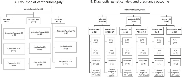
Objective: To investigate the clinical outcome of fetuses with ventriculomegaly (VM), and to identify risk factors for progression of fetal VM in order to improve prenatal counseling. This was a multicenter, retrospective cohort study, comprising 229 cases with VM.
Methods: VM was classified as mild, moderate, or severe and isolated or non-isolated. Genetic data were collected. Differences between VM subgroups were described, and risk factors for progression of fetal VM were identified using logistic regression analysis. Outcome was defined as the percentage of live births, termination of pregnancy (TOP) and intra-uterine fetal demise (IUFD).
Results: Of the 229 cases, 109 (47.6%) had mild VM, 60 (26.2%) moderate VM, and 60 (26.2%) severe VM. Progression of VM occurred in 45/153 cases (29.4%), half of which were in the group with severe VM. Dilatation of the 3rd ventricle and neural tube defects were risk factors for progression of VM. The percentage of live births (excluding cases with TOP and unknown outcome) was 93.1% (54/58) in mild VM, 78.6% (22/28) in moderate VM and 92.6% (25/27) in severe VM. In 12/229 cases (5.2%) IUFD occurred. Genetic analysis was performed in 143/229 (62.4%) of cases, showing (likely) pathogenic abnormalities in 41/143 (28.7%) cases, predominantly in mild, non-isolated VM.
Conclusions: This study confirms the clinical relevance of additional genetic investigations in all types of fetal VMs. Further larger prospective research including clinical follow-up is needed to improve prenatal counseling.
期刊介绍:
Prenatal Diagnosis welcomes submissions in all aspects of prenatal diagnosis with a particular focus on areas in which molecular biology and genetics interface with prenatal care and therapy, encompassing: all aspects of fetal imaging, including sonography and magnetic resonance imaging; prenatal cytogenetics, including molecular studies and array CGH; prenatal screening studies; fetal cells and cell-free nucleic acids in maternal blood and other fluids; preimplantation genetic diagnosis (PGD); prenatal diagnosis of single gene disorders, including metabolic disorders; fetal therapy; fetal and placental development and pathology; development and evaluation of laboratory services for prenatal diagnosis; psychosocial, legal, ethical and economic aspects of prenatal diagnosis; prenatal genetic counseling
 求助内容:
求助内容: 应助结果提醒方式:


