Monder Lafi, Hamzah Amin, Muhammed Aqib Khan, Marwan Bukhari
{"title":"三分之一桡骨骨密度测量在骨质疏松症诊断中的应用:与股骨和腰椎骨密度的比较分析。","authors":"Monder Lafi, Hamzah Amin, Muhammed Aqib Khan, Marwan Bukhari","doi":"10.1080/19932820.2025.2506877","DOIUrl":null,"url":null,"abstract":"<p><p>Osteoporosis is defined by a BMD ≤ 2.5 SD below the young adult reference population. Standard dual-energy X-ray absorptiometry (DXA) scans for osteoporosis involve the femoral neck and lumbar spine, but alternative sites like the one-third radius (1/3 R) are only used when these sites are inaccessible. This study assessed the correlation and level of agreement between BMD at the 1/3 R, femoral neck, and lumbar spine to evaluate its diagnostic utility. Data from 43,801 patients referred for DXA scans in northwest England were analysed. Of these, 437 underwent 1/3 R scans. Demographic comparisons between patients with and without forearm scans were conducted. The primary analysis included patients with scans at the 1/3 R, lumbar spine, and bilateral femoral regions;(<i>n</i> = 183). Spearman's correlation assessed BMD relationships, Cohen's kappa analysed osteoporosis classification agreement, and Bland-Altman plots evaluated measurement bias. The cohort had a mean age of 65.7 years (SD 12.9), with 83.3% female and 41.2% reporting fractures. Patients who underwent 1/3 R scans (<i>n</i> = 437) were older, heavier, and had a higher body mass index (BMI). Correlation analysis showed only moderate associations between 1/3 R and femoral/lumbar spine BMD ;(<i>r</i> = 0.29 to 0.36, <i>p</i> < 0.001). Cohen's kappa demonstrated only slight agreement for 1/3 R, femoral neck and lumbar spine T-scores (κ = 0.14-0.29). Bland-Altman analysis revealed that 1/3 R scans systematically underestimated BMD relative to femoral and lumbar sites, with mean biases of -0.7 for femoral sites and -1.53 for lumbar spine. The 1/3 R BMD showed poor agreement and systematic underestimation compared to central sites, limiting its reliability for osteoporosis diagnosis. Future research should explore alternative peripheral weight-bearing sites and novel diagnostic technologies to assess BMD where central sites cannot be scanned.</p>","PeriodicalId":49910,"journal":{"name":"Libyan Journal of Medicine","volume":"20 1","pages":"2506877"},"PeriodicalIF":1.7000,"publicationDate":"2025-12-01","publicationTypes":"Journal Article","fieldsOfStudy":null,"isOpenAccess":false,"openAccessPdf":"https://www.ncbi.nlm.nih.gov/pmc/articles/PMC12086901/pdf/","citationCount":"0","resultStr":"{\"title\":\"One-third Radius bone mineral density measurement utility in the diagnosis of osteoporosis: a comparative analysis with femoral and lumbar spine bone mineral density.\",\"authors\":\"Monder Lafi, Hamzah Amin, Muhammed Aqib Khan, Marwan Bukhari\",\"doi\":\"10.1080/19932820.2025.2506877\",\"DOIUrl\":null,\"url\":null,\"abstract\":\"<p><p>Osteoporosis is defined by a BMD ≤ 2.5 SD below the young adult reference population. Standard dual-energy X-ray absorptiometry (DXA) scans for osteoporosis involve the femoral neck and lumbar spine, but alternative sites like the one-third radius (1/3 R) are only used when these sites are inaccessible. This study assessed the correlation and level of agreement between BMD at the 1/3 R, femoral neck, and lumbar spine to evaluate its diagnostic utility. Data from 43,801 patients referred for DXA scans in northwest England were analysed. Of these, 437 underwent 1/3 R scans. Demographic comparisons between patients with and without forearm scans were conducted. The primary analysis included patients with scans at the 1/3 R, lumbar spine, and bilateral femoral regions;(<i>n</i> = 183). Spearman's correlation assessed BMD relationships, Cohen's kappa analysed osteoporosis classification agreement, and Bland-Altman plots evaluated measurement bias. The cohort had a mean age of 65.7 years (SD 12.9), with 83.3% female and 41.2% reporting fractures. Patients who underwent 1/3 R scans (<i>n</i> = 437) were older, heavier, and had a higher body mass index (BMI). Correlation analysis showed only moderate associations between 1/3 R and femoral/lumbar spine BMD ;(<i>r</i> = 0.29 to 0.36, <i>p</i> < 0.001). Cohen's kappa demonstrated only slight agreement for 1/3 R, femoral neck and lumbar spine T-scores (κ = 0.14-0.29). Bland-Altman analysis revealed that 1/3 R scans systematically underestimated BMD relative to femoral and lumbar sites, with mean biases of -0.7 for femoral sites and -1.53 for lumbar spine. The 1/3 R BMD showed poor agreement and systematic underestimation compared to central sites, limiting its reliability for osteoporosis diagnosis. Future research should explore alternative peripheral weight-bearing sites and novel diagnostic technologies to assess BMD where central sites cannot be scanned.</p>\",\"PeriodicalId\":49910,\"journal\":{\"name\":\"Libyan Journal of Medicine\",\"volume\":\"20 1\",\"pages\":\"2506877\"},\"PeriodicalIF\":1.7000,\"publicationDate\":\"2025-12-01\",\"publicationTypes\":\"Journal Article\",\"fieldsOfStudy\":null,\"isOpenAccess\":false,\"openAccessPdf\":\"https://www.ncbi.nlm.nih.gov/pmc/articles/PMC12086901/pdf/\",\"citationCount\":\"0\",\"resultStr\":null,\"platform\":\"Semanticscholar\",\"paperid\":null,\"PeriodicalName\":\"Libyan Journal of Medicine\",\"FirstCategoryId\":\"3\",\"ListUrlMain\":\"https://doi.org/10.1080/19932820.2025.2506877\",\"RegionNum\":4,\"RegionCategory\":\"医学\",\"ArticlePicture\":[],\"TitleCN\":null,\"AbstractTextCN\":null,\"PMCID\":null,\"EPubDate\":\"2025/5/17 0:00:00\",\"PubModel\":\"Epub\",\"JCR\":\"Q2\",\"JCRName\":\"MEDICINE, GENERAL & INTERNAL\",\"Score\":null,\"Total\":0}","platform":"Semanticscholar","paperid":null,"PeriodicalName":"Libyan Journal of Medicine","FirstCategoryId":"3","ListUrlMain":"https://doi.org/10.1080/19932820.2025.2506877","RegionNum":4,"RegionCategory":"医学","ArticlePicture":[],"TitleCN":null,"AbstractTextCN":null,"PMCID":null,"EPubDate":"2025/5/17 0:00:00","PubModel":"Epub","JCR":"Q2","JCRName":"MEDICINE, GENERAL & INTERNAL","Score":null,"Total":0}
One-third Radius bone mineral density measurement utility in the diagnosis of osteoporosis: a comparative analysis with femoral and lumbar spine bone mineral density.
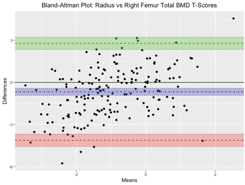
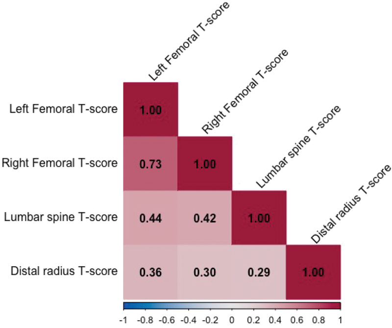
Osteoporosis is defined by a BMD ≤ 2.5 SD below the young adult reference population. Standard dual-energy X-ray absorptiometry (DXA) scans for osteoporosis involve the femoral neck and lumbar spine, but alternative sites like the one-third radius (1/3 R) are only used when these sites are inaccessible. This study assessed the correlation and level of agreement between BMD at the 1/3 R, femoral neck, and lumbar spine to evaluate its diagnostic utility. Data from 43,801 patients referred for DXA scans in northwest England were analysed. Of these, 437 underwent 1/3 R scans. Demographic comparisons between patients with and without forearm scans were conducted. The primary analysis included patients with scans at the 1/3 R, lumbar spine, and bilateral femoral regions;(n = 183). Spearman's correlation assessed BMD relationships, Cohen's kappa analysed osteoporosis classification agreement, and Bland-Altman plots evaluated measurement bias. The cohort had a mean age of 65.7 years (SD 12.9), with 83.3% female and 41.2% reporting fractures. Patients who underwent 1/3 R scans (n = 437) were older, heavier, and had a higher body mass index (BMI). Correlation analysis showed only moderate associations between 1/3 R and femoral/lumbar spine BMD ;(r = 0.29 to 0.36, p < 0.001). Cohen's kappa demonstrated only slight agreement for 1/3 R, femoral neck and lumbar spine T-scores (κ = 0.14-0.29). Bland-Altman analysis revealed that 1/3 R scans systematically underestimated BMD relative to femoral and lumbar sites, with mean biases of -0.7 for femoral sites and -1.53 for lumbar spine. The 1/3 R BMD showed poor agreement and systematic underestimation compared to central sites, limiting its reliability for osteoporosis diagnosis. Future research should explore alternative peripheral weight-bearing sites and novel diagnostic technologies to assess BMD where central sites cannot be scanned.
期刊介绍:
Libyan Journal of Medicine (LJM) is a peer-reviewed, Open Access, international medical journal aiming to promote heath and health education by publishing high-quality medical research in the different disciplines of medicine.
LJM was founded in 2006 by a group of enthusiastic Libyan medical scientists who looked at the contribution of Libyan publications to the international medical literature and saw that a publication outlet was missing. To fill this gap they launched LJM as a tool for transferring current medical knowledge to and from colleagues in developing countries, particularly African countries, as well as internationally.The journal is still led by a group of Libyan physicians inside and outside Libya, but it also enjoys support and recognition from the international medical community.


 求助内容:
求助内容: 应助结果提醒方式:


