Shanshan Liu, Chaoqun Han, Qi He, Guochen Shang, Yu Jin, Jun Liu, Zhen Ding, Rong Lin
{"title":"胃印戒细胞癌两种不同表现的临床病理特征比较。","authors":"Shanshan Liu, Chaoqun Han, Qi He, Guochen Shang, Yu Jin, Jun Liu, Zhen Ding, Rong Lin","doi":"10.1097/eus.0000000000000085","DOIUrl":null,"url":null,"abstract":"<p><strong>Background and objectives: </strong>There are two different endoscopic ultrasonographic manifestations of gastric signet ring cell carcinoma (GSRCC). No studies have been reported on the differences in the clinical profiles of patients based on EUS examination. We aim to study the variations in clinicopathological characteristics between two distinct endoscopic ultrasonographic manifestations of GSRCC.</p><p><strong>Methods: </strong>A total of 302 patients with GSRCC confirmed by pathological examination who underwent EUS were enrolled in the study. Based on the endoscopic ultrasonographic features, patients were categorized into two groups: type 1, where the entire layer structure disappeared, and type 2, where the layer structure was still present and appeared lymphomatoid. Clinicopathologic features were collected retrospectively and analyzed.</p><p><strong>Results: </strong>Compared with type 2 patients, type 1 patients tended to develop GSRCC at an older age (<i>P</i> = 0.033) and had higher serum levels of tumor markers and were more likely to experience anemia (<i>P</i> < 0.001) and weight loss (<i>P</i> < 0.001) during the disease progression. Significant increases in the tumor size (<i>P</i> < 0.001), thickness of the affected gastric wall (<i>P</i> < 0.001), and depth of tumor invasion (<i>P</i> < 0.001) were observed in type 1 patients. Furthermore, type 1 patients had higher prevalence of affected blood vessels (<i>P</i> < 0.001), nerves (<i>P</i> < 0.001), lymph nodes (<i>P</i> < 0.001), and peritoneal metastasis (<i>P</i> < 0.001). However, no difference was found in the duration of disease between the two groups, and all deficient mismatch repairs were observed in type 1 patients.</p><p><strong>Conclusions: </strong>The two distinct endoscopic ultrasonographic manifestations of GSRCC exhibited different clinicopathological characteristics, suggesting that they may represent different subtypes of the disease that require special attention in management strategies.</p>","PeriodicalId":11577,"journal":{"name":"Endoscopic Ultrasound","volume":"13 5","pages":"293-299"},"PeriodicalIF":5.4000,"publicationDate":"2024-09-01","publicationTypes":"Journal Article","fieldsOfStudy":null,"isOpenAccess":false,"openAccessPdf":"https://www.ncbi.nlm.nih.gov/pmc/articles/PMC12080688/pdf/","citationCount":"0","resultStr":"{\"title\":\"The comparison of clinicopathological characteristics of two distinct manifestations of gastric signet ring cell carcinoma under EUS.\",\"authors\":\"Shanshan Liu, Chaoqun Han, Qi He, Guochen Shang, Yu Jin, Jun Liu, Zhen Ding, Rong Lin\",\"doi\":\"10.1097/eus.0000000000000085\",\"DOIUrl\":null,\"url\":null,\"abstract\":\"<p><strong>Background and objectives: </strong>There are two different endoscopic ultrasonographic manifestations of gastric signet ring cell carcinoma (GSRCC). No studies have been reported on the differences in the clinical profiles of patients based on EUS examination. We aim to study the variations in clinicopathological characteristics between two distinct endoscopic ultrasonographic manifestations of GSRCC.</p><p><strong>Methods: </strong>A total of 302 patients with GSRCC confirmed by pathological examination who underwent EUS were enrolled in the study. Based on the endoscopic ultrasonographic features, patients were categorized into two groups: type 1, where the entire layer structure disappeared, and type 2, where the layer structure was still present and appeared lymphomatoid. Clinicopathologic features were collected retrospectively and analyzed.</p><p><strong>Results: </strong>Compared with type 2 patients, type 1 patients tended to develop GSRCC at an older age (<i>P</i> = 0.033) and had higher serum levels of tumor markers and were more likely to experience anemia (<i>P</i> < 0.001) and weight loss (<i>P</i> < 0.001) during the disease progression. Significant increases in the tumor size (<i>P</i> < 0.001), thickness of the affected gastric wall (<i>P</i> < 0.001), and depth of tumor invasion (<i>P</i> < 0.001) were observed in type 1 patients. Furthermore, type 1 patients had higher prevalence of affected blood vessels (<i>P</i> < 0.001), nerves (<i>P</i> < 0.001), lymph nodes (<i>P</i> < 0.001), and peritoneal metastasis (<i>P</i> < 0.001). However, no difference was found in the duration of disease between the two groups, and all deficient mismatch repairs were observed in type 1 patients.</p><p><strong>Conclusions: </strong>The two distinct endoscopic ultrasonographic manifestations of GSRCC exhibited different clinicopathological characteristics, suggesting that they may represent different subtypes of the disease that require special attention in management strategies.</p>\",\"PeriodicalId\":11577,\"journal\":{\"name\":\"Endoscopic Ultrasound\",\"volume\":\"13 5\",\"pages\":\"293-299\"},\"PeriodicalIF\":5.4000,\"publicationDate\":\"2024-09-01\",\"publicationTypes\":\"Journal Article\",\"fieldsOfStudy\":null,\"isOpenAccess\":false,\"openAccessPdf\":\"https://www.ncbi.nlm.nih.gov/pmc/articles/PMC12080688/pdf/\",\"citationCount\":\"0\",\"resultStr\":null,\"platform\":\"Semanticscholar\",\"paperid\":null,\"PeriodicalName\":\"Endoscopic Ultrasound\",\"FirstCategoryId\":\"3\",\"ListUrlMain\":\"https://doi.org/10.1097/eus.0000000000000085\",\"RegionNum\":1,\"RegionCategory\":\"医学\",\"ArticlePicture\":[],\"TitleCN\":null,\"AbstractTextCN\":null,\"PMCID\":null,\"EPubDate\":\"2024/11/8 0:00:00\",\"PubModel\":\"Epub\",\"JCR\":\"Q1\",\"JCRName\":\"GASTROENTEROLOGY & HEPATOLOGY\",\"Score\":null,\"Total\":0}","platform":"Semanticscholar","paperid":null,"PeriodicalName":"Endoscopic Ultrasound","FirstCategoryId":"3","ListUrlMain":"https://doi.org/10.1097/eus.0000000000000085","RegionNum":1,"RegionCategory":"医学","ArticlePicture":[],"TitleCN":null,"AbstractTextCN":null,"PMCID":null,"EPubDate":"2024/11/8 0:00:00","PubModel":"Epub","JCR":"Q1","JCRName":"GASTROENTEROLOGY & HEPATOLOGY","Score":null,"Total":0}
The comparison of clinicopathological characteristics of two distinct manifestations of gastric signet ring cell carcinoma under EUS.
Background and objectives: There are two different endoscopic ultrasonographic manifestations of gastric signet ring cell carcinoma (GSRCC). No studies have been reported on the differences in the clinical profiles of patients based on EUS examination. We aim to study the variations in clinicopathological characteristics between two distinct endoscopic ultrasonographic manifestations of GSRCC.
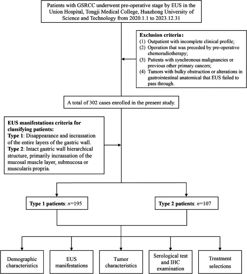
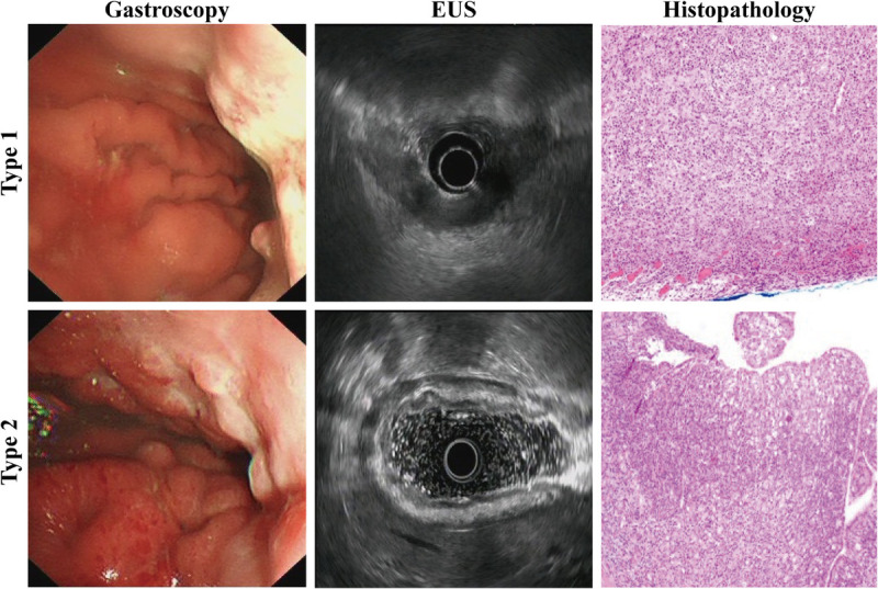
Methods: A total of 302 patients with GSRCC confirmed by pathological examination who underwent EUS were enrolled in the study. Based on the endoscopic ultrasonographic features, patients were categorized into two groups: type 1, where the entire layer structure disappeared, and type 2, where the layer structure was still present and appeared lymphomatoid. Clinicopathologic features were collected retrospectively and analyzed.
Results: Compared with type 2 patients, type 1 patients tended to develop GSRCC at an older age (P = 0.033) and had higher serum levels of tumor markers and were more likely to experience anemia (P < 0.001) and weight loss (P < 0.001) during the disease progression. Significant increases in the tumor size (P < 0.001), thickness of the affected gastric wall (P < 0.001), and depth of tumor invasion (P < 0.001) were observed in type 1 patients. Furthermore, type 1 patients had higher prevalence of affected blood vessels (P < 0.001), nerves (P < 0.001), lymph nodes (P < 0.001), and peritoneal metastasis (P < 0.001). However, no difference was found in the duration of disease between the two groups, and all deficient mismatch repairs were observed in type 1 patients.
Conclusions: The two distinct endoscopic ultrasonographic manifestations of GSRCC exhibited different clinicopathological characteristics, suggesting that they may represent different subtypes of the disease that require special attention in management strategies.
期刊介绍:
Endoscopic Ultrasound, a publication of Euro-EUS Scientific Committee, Asia-Pacific EUS Task Force and Latin American Chapter of EUS, is a peer-reviewed online journal with Quarterly print on demand compilation of issues published. The journal’s full text is available online at http://www.eusjournal.com. The journal allows free access (Open Access) to its contents and permits authors to self-archive final accepted version of the articles on any OAI-compliant institutional / subject-based repository. The journal does not charge for submission, processing or publication of manuscripts and even for color reproduction of photographs.
 求助内容:
求助内容: 应助结果提醒方式:


