Stephen S Adler, Noriko Sato, Kwamena E Baidoo, Frank I Lin, Woonghee Lee, Colleen P Olkowski, Freddy E Escorcia, Peter L Choyke
{"title":"一种定量粒子识别(QPID)光谱放射自成像系统。","authors":"Stephen S Adler, Noriko Sato, Kwamena E Baidoo, Frank I Lin, Woonghee Lee, Colleen P Olkowski, Freddy E Escorcia, Peter L Choyke","doi":"10.1038/s44172-025-00426-1","DOIUrl":null,"url":null,"abstract":"<p><p>Autoradiography is used to study the distribution and binding of radioisotope tagged ligands in tissue at microscale among other applications. The technology has evolved since its inception when it used analogue film exposure techniques with the introduction of digital imaging systems sensitive to ionizing radiation. We report on the development of our Quantitative Particle Identification spectral autoradiography system (QPID), which is based on the Timepix3 sensor and a gamma detecting scintillation crystal. This autoradiography system leverages the ionizing radiation detection features of the Timepix3 to measure the energy deposition from charged particles from radioisotopes with a time resolution of 7.7 ns full width at half max (FWHM), generating spectral or activity autoradiography images. The QPID includes a scintillation crystal used to record gamma emissions coincident with the Timepix3 ionization events with a time resolution of 24.2 ns FWHM. The QPID can separate tracks between α and ß particles, select specific ranges of deposited energies or select on the presence of coincident gamma emissions within a selected energy range when generating images. The QPID has a 10% linearity response up to 700 Bq for 223Ra and 2.5 kBq for 18 F radioisotopes. Using α and ß+ particle identification filters, separate images of <sup>223</sup>RaCl<sub>2</sub> and Na<sup>18</sup>F activity distributions were generated from a bone sample infused with the two radioligands together. This unique capability can open the door to the study of targeted radiotherapies which use theranostic α and ß+ imaging agents by measuring their relative pharmacokinetic properties.</p>","PeriodicalId":72644,"journal":{"name":"Communications engineering","volume":"4 1","pages":"89"},"PeriodicalIF":0.0000,"publicationDate":"2025-05-15","publicationTypes":"Journal Article","fieldsOfStudy":null,"isOpenAccess":false,"openAccessPdf":"https://www.ncbi.nlm.nih.gov/pmc/articles/PMC12081844/pdf/","citationCount":"0","resultStr":"{\"title\":\"A Quantitative Particle Identification (QPID) spectral autoradiography system.\",\"authors\":\"Stephen S Adler, Noriko Sato, Kwamena E Baidoo, Frank I Lin, Woonghee Lee, Colleen P Olkowski, Freddy E Escorcia, Peter L Choyke\",\"doi\":\"10.1038/s44172-025-00426-1\",\"DOIUrl\":null,\"url\":null,\"abstract\":\"<p><p>Autoradiography is used to study the distribution and binding of radioisotope tagged ligands in tissue at microscale among other applications. The technology has evolved since its inception when it used analogue film exposure techniques with the introduction of digital imaging systems sensitive to ionizing radiation. We report on the development of our Quantitative Particle Identification spectral autoradiography system (QPID), which is based on the Timepix3 sensor and a gamma detecting scintillation crystal. This autoradiography system leverages the ionizing radiation detection features of the Timepix3 to measure the energy deposition from charged particles from radioisotopes with a time resolution of 7.7 ns full width at half max (FWHM), generating spectral or activity autoradiography images. The QPID includes a scintillation crystal used to record gamma emissions coincident with the Timepix3 ionization events with a time resolution of 24.2 ns FWHM. The QPID can separate tracks between α and ß particles, select specific ranges of deposited energies or select on the presence of coincident gamma emissions within a selected energy range when generating images. The QPID has a 10% linearity response up to 700 Bq for 223Ra and 2.5 kBq for 18 F radioisotopes. Using α and ß+ particle identification filters, separate images of <sup>223</sup>RaCl<sub>2</sub> and Na<sup>18</sup>F activity distributions were generated from a bone sample infused with the two radioligands together. This unique capability can open the door to the study of targeted radiotherapies which use theranostic α and ß+ imaging agents by measuring their relative pharmacokinetic properties.</p>\",\"PeriodicalId\":72644,\"journal\":{\"name\":\"Communications engineering\",\"volume\":\"4 1\",\"pages\":\"89\"},\"PeriodicalIF\":0.0000,\"publicationDate\":\"2025-05-15\",\"publicationTypes\":\"Journal Article\",\"fieldsOfStudy\":null,\"isOpenAccess\":false,\"openAccessPdf\":\"https://www.ncbi.nlm.nih.gov/pmc/articles/PMC12081844/pdf/\",\"citationCount\":\"0\",\"resultStr\":null,\"platform\":\"Semanticscholar\",\"paperid\":null,\"PeriodicalName\":\"Communications engineering\",\"FirstCategoryId\":\"1085\",\"ListUrlMain\":\"https://doi.org/10.1038/s44172-025-00426-1\",\"RegionNum\":0,\"RegionCategory\":null,\"ArticlePicture\":[],\"TitleCN\":null,\"AbstractTextCN\":null,\"PMCID\":null,\"EPubDate\":\"\",\"PubModel\":\"\",\"JCR\":\"\",\"JCRName\":\"\",\"Score\":null,\"Total\":0}","platform":"Semanticscholar","paperid":null,"PeriodicalName":"Communications engineering","FirstCategoryId":"1085","ListUrlMain":"https://doi.org/10.1038/s44172-025-00426-1","RegionNum":0,"RegionCategory":null,"ArticlePicture":[],"TitleCN":null,"AbstractTextCN":null,"PMCID":null,"EPubDate":"","PubModel":"","JCR":"","JCRName":"","Score":null,"Total":0}
A Quantitative Particle Identification (QPID) spectral autoradiography system.
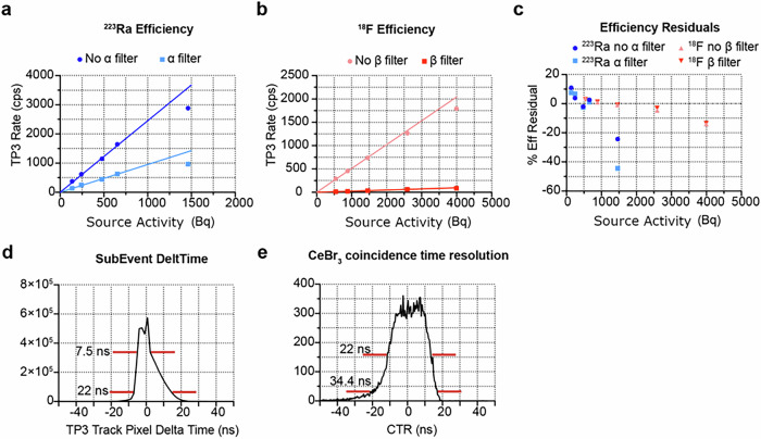
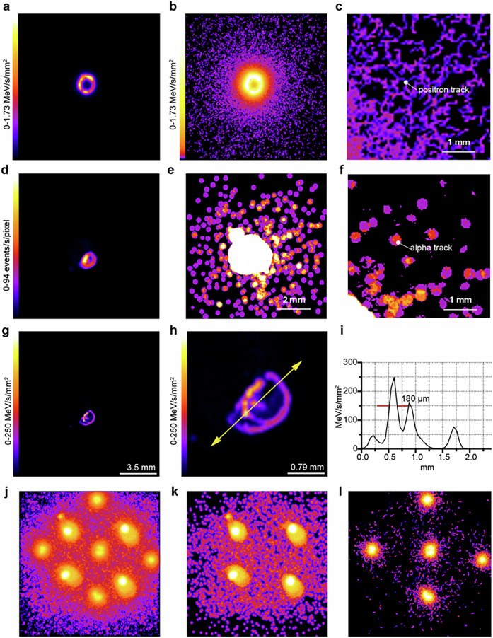
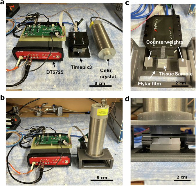
Autoradiography is used to study the distribution and binding of radioisotope tagged ligands in tissue at microscale among other applications. The technology has evolved since its inception when it used analogue film exposure techniques with the introduction of digital imaging systems sensitive to ionizing radiation. We report on the development of our Quantitative Particle Identification spectral autoradiography system (QPID), which is based on the Timepix3 sensor and a gamma detecting scintillation crystal. This autoradiography system leverages the ionizing radiation detection features of the Timepix3 to measure the energy deposition from charged particles from radioisotopes with a time resolution of 7.7 ns full width at half max (FWHM), generating spectral or activity autoradiography images. The QPID includes a scintillation crystal used to record gamma emissions coincident with the Timepix3 ionization events with a time resolution of 24.2 ns FWHM. The QPID can separate tracks between α and ß particles, select specific ranges of deposited energies or select on the presence of coincident gamma emissions within a selected energy range when generating images. The QPID has a 10% linearity response up to 700 Bq for 223Ra and 2.5 kBq for 18 F radioisotopes. Using α and ß+ particle identification filters, separate images of 223RaCl2 and Na18F activity distributions were generated from a bone sample infused with the two radioligands together. This unique capability can open the door to the study of targeted radiotherapies which use theranostic α and ß+ imaging agents by measuring their relative pharmacokinetic properties.

 求助内容:
求助内容: 应助结果提醒方式:


