{"title":"无病受试者脊髓18F-FDG的生理分布。","authors":"Selin Kesim, Salih Özgüven","doi":"10.4274/mirt.galenos.2025.56514","DOIUrl":null,"url":null,"abstract":"<p><strong>Objectives: </strong><sup>18</sup>Fluorine-fluorodeoxyglucose (<sup>18</sup>F-FDG) uptake in the spinal cord is not unusual and may mimic metastatic disease. The physiological characterization and variability of spinal cord <sup>18</sup>F-FDG metabolism provide valuable information, especially in patients with suspected malignancies. We aimed to investigate the physiological <sup>18</sup>F-FDG uptake pattern within the spinal cord and its associations in a normal population.</p><p><strong>Methods: </strong>We retrospectively analyzed <sup>18</sup>F-FDG positron emission tomography/computed tomography images of 140 adult patients who were confirmed to be disease-free over a one-year follow-up period. The maximal and mean standard uptake values (SUV<sub>max</sub>, SUV<sub>mean</sub>) were measured at each mid-vertebral level from C1 to L5, and normalized to liver and blood pool uptake. Correlations between <sup>18</sup>F-FDG uptake and patient demographics, clinical parameters, and environmental temperature were evaluated.</p><p><strong>Results: </strong><sup>18</sup>F-FDG uptake demonstrated a decreasing trend from the cervical to lumbar vertebrae, with a notable increase at the lower thoracic levels (T11-T12). There was a significant negative correlation between <sup>18</sup>F-FDG uptake and age (p<0.001), fasting glucose level (p=0.016), and diabetic status (p=0.003). No significant association was found between spinal cord <sup>18</sup>F-FDG uptake and gender, weight, height, body mass index, <sup>18</sup>F-FDG dose, or environmental temperature.</p><p><strong>Conclusion: </strong>Normal distribution of <sup>18</sup>F-FDG in the spinal cord of disease-free individuals decreases from cervical to lumbar levels, although it notably increases at the lower thoracic and mid-lower cervical levels. Uptake significantly decreases with age, with a higher fasting blood glucose level, and in diabetic patients.</p>","PeriodicalId":44681,"journal":{"name":"Molecular Imaging and Radionuclide Therapy","volume":" ","pages":"107-113"},"PeriodicalIF":1.1000,"publicationDate":"2025-06-03","publicationTypes":"Journal Article","fieldsOfStudy":null,"isOpenAccess":false,"openAccessPdf":"https://www.ncbi.nlm.nih.gov/pmc/articles/PMC12134945/pdf/","citationCount":"0","resultStr":"{\"title\":\"Physiological Distribution of <sup>18</sup>F-FDG in the Spinal Cord of Disease-Free Subjects\",\"authors\":\"Selin Kesim, Salih Özgüven\",\"doi\":\"10.4274/mirt.galenos.2025.56514\",\"DOIUrl\":null,\"url\":null,\"abstract\":\"<p><strong>Objectives: </strong><sup>18</sup>Fluorine-fluorodeoxyglucose (<sup>18</sup>F-FDG) uptake in the spinal cord is not unusual and may mimic metastatic disease. The physiological characterization and variability of spinal cord <sup>18</sup>F-FDG metabolism provide valuable information, especially in patients with suspected malignancies. We aimed to investigate the physiological <sup>18</sup>F-FDG uptake pattern within the spinal cord and its associations in a normal population.</p><p><strong>Methods: </strong>We retrospectively analyzed <sup>18</sup>F-FDG positron emission tomography/computed tomography images of 140 adult patients who were confirmed to be disease-free over a one-year follow-up period. The maximal and mean standard uptake values (SUV<sub>max</sub>, SUV<sub>mean</sub>) were measured at each mid-vertebral level from C1 to L5, and normalized to liver and blood pool uptake. Correlations between <sup>18</sup>F-FDG uptake and patient demographics, clinical parameters, and environmental temperature were evaluated.</p><p><strong>Results: </strong><sup>18</sup>F-FDG uptake demonstrated a decreasing trend from the cervical to lumbar vertebrae, with a notable increase at the lower thoracic levels (T11-T12). There was a significant negative correlation between <sup>18</sup>F-FDG uptake and age (p<0.001), fasting glucose level (p=0.016), and diabetic status (p=0.003). No significant association was found between spinal cord <sup>18</sup>F-FDG uptake and gender, weight, height, body mass index, <sup>18</sup>F-FDG dose, or environmental temperature.</p><p><strong>Conclusion: </strong>Normal distribution of <sup>18</sup>F-FDG in the spinal cord of disease-free individuals decreases from cervical to lumbar levels, although it notably increases at the lower thoracic and mid-lower cervical levels. Uptake significantly decreases with age, with a higher fasting blood glucose level, and in diabetic patients.</p>\",\"PeriodicalId\":44681,\"journal\":{\"name\":\"Molecular Imaging and Radionuclide Therapy\",\"volume\":\" \",\"pages\":\"107-113\"},\"PeriodicalIF\":1.1000,\"publicationDate\":\"2025-06-03\",\"publicationTypes\":\"Journal Article\",\"fieldsOfStudy\":null,\"isOpenAccess\":false,\"openAccessPdf\":\"https://www.ncbi.nlm.nih.gov/pmc/articles/PMC12134945/pdf/\",\"citationCount\":\"0\",\"resultStr\":null,\"platform\":\"Semanticscholar\",\"paperid\":null,\"PeriodicalName\":\"Molecular Imaging and Radionuclide Therapy\",\"FirstCategoryId\":\"1085\",\"ListUrlMain\":\"https://doi.org/10.4274/mirt.galenos.2025.56514\",\"RegionNum\":0,\"RegionCategory\":null,\"ArticlePicture\":[],\"TitleCN\":null,\"AbstractTextCN\":null,\"PMCID\":null,\"EPubDate\":\"2025/5/15 0:00:00\",\"PubModel\":\"Epub\",\"JCR\":\"Q4\",\"JCRName\":\"RADIOLOGY, NUCLEAR MEDICINE & MEDICAL IMAGING\",\"Score\":null,\"Total\":0}","platform":"Semanticscholar","paperid":null,"PeriodicalName":"Molecular Imaging and Radionuclide Therapy","FirstCategoryId":"1085","ListUrlMain":"https://doi.org/10.4274/mirt.galenos.2025.56514","RegionNum":0,"RegionCategory":null,"ArticlePicture":[],"TitleCN":null,"AbstractTextCN":null,"PMCID":null,"EPubDate":"2025/5/15 0:00:00","PubModel":"Epub","JCR":"Q4","JCRName":"RADIOLOGY, NUCLEAR MEDICINE & MEDICAL IMAGING","Score":null,"Total":0}
Physiological Distribution of 18F-FDG in the Spinal Cord of Disease-Free Subjects
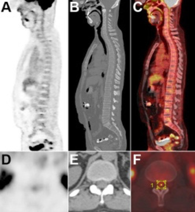
Objectives: 18Fluorine-fluorodeoxyglucose (18F-FDG) uptake in the spinal cord is not unusual and may mimic metastatic disease. The physiological characterization and variability of spinal cord 18F-FDG metabolism provide valuable information, especially in patients with suspected malignancies. We aimed to investigate the physiological 18F-FDG uptake pattern within the spinal cord and its associations in a normal population.
Methods: We retrospectively analyzed 18F-FDG positron emission tomography/computed tomography images of 140 adult patients who were confirmed to be disease-free over a one-year follow-up period. The maximal and mean standard uptake values (SUVmax, SUVmean) were measured at each mid-vertebral level from C1 to L5, and normalized to liver and blood pool uptake. Correlations between 18F-FDG uptake and patient demographics, clinical parameters, and environmental temperature were evaluated.
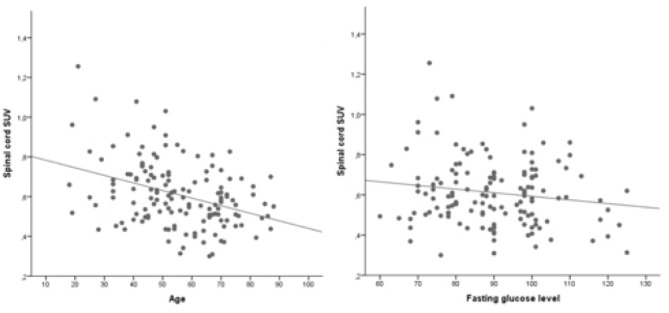
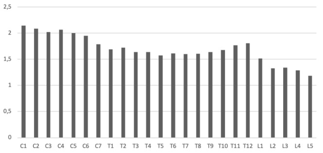
Results: 18F-FDG uptake demonstrated a decreasing trend from the cervical to lumbar vertebrae, with a notable increase at the lower thoracic levels (T11-T12). There was a significant negative correlation between 18F-FDG uptake and age (p<0.001), fasting glucose level (p=0.016), and diabetic status (p=0.003). No significant association was found between spinal cord 18F-FDG uptake and gender, weight, height, body mass index, 18F-FDG dose, or environmental temperature.
Conclusion: Normal distribution of 18F-FDG in the spinal cord of disease-free individuals decreases from cervical to lumbar levels, although it notably increases at the lower thoracic and mid-lower cervical levels. Uptake significantly decreases with age, with a higher fasting blood glucose level, and in diabetic patients.
期刊介绍:
Molecular Imaging and Radionuclide Therapy (Mol Imaging Radionucl Ther, MIRT) is publishes original research articles, invited reviews, editorials, short communications, letters, consensus statements, guidelines and case reports with a literature review on the topic, in the field of molecular imaging, multimodality imaging, nuclear medicine, radionuclide therapy, radiopharmacy, medical physics, dosimetry and radiobiology.

 求助内容:
求助内容: 应助结果提醒方式:


