Rubens Peres Mendes, Aymara Eduarda de Lima, Reginaldo da Cunha, Mauricio Jose Bittar, Christian Carlstron Vasconcelos, Diego Darley Velasquez Piñeros, Rodrigo Romero Corrêa
{"title":"互补放射线投影评价马鼻窦和大泡。","authors":"Rubens Peres Mendes, Aymara Eduarda de Lima, Reginaldo da Cunha, Mauricio Jose Bittar, Christian Carlstron Vasconcelos, Diego Darley Velasquez Piñeros, Rodrigo Romero Corrêa","doi":"10.1111/vru.70046","DOIUrl":null,"url":null,"abstract":"<p><p>Radiographic examination of the skull is a well-established and indispensable procedure for the diagnostic evaluation of dental and paranasal sinus disorders in horses. Complementary projections make significant contributions to radiographic diagnosis in nasal conchae disorders. This article describes a complementary radiographic projection designed for the evaluation of the conchal sinuses and bullae in horses. Six equine cadaveric heads were used. Specimens were dissected, and openings were created in the ventral and dorsal nasal conchae for the introduction of radiopaque material. The new radiographic projection was named lateral 75° dorsal-lateral ventral oblique view. This projection was obtained with the mandibular rami resting on the imaging plate and the mandible displaced toward the side of interest (partial excursion). The X-ray beam was directed dorsoventrally, slightly angled toward the side of interest (left or right offset mandible dorsoventral view), and centered at an imaginary line connecting the tips of the facial crests. The lateral 75° dorsal-lateral ventral oblique view provided enhanced visualization and allowed correct identification of equine conchal structures. Lateral displacement of the mandible and the 75° angle of inclination to the vertical plane eliminated the superimposition of anatomical structures, facilitating radiographic image interpretation and increasing diagnostic accuracy. This complementary projection is recommended in all cases of sinonasal disorders in horses.</p>","PeriodicalId":23581,"journal":{"name":"Veterinary Radiology & Ultrasound","volume":"66 3","pages":"e70046"},"PeriodicalIF":1.5000,"publicationDate":"2025-05-01","publicationTypes":"Journal Article","fieldsOfStudy":null,"isOpenAccess":false,"openAccessPdf":"https://www.ncbi.nlm.nih.gov/pmc/articles/PMC12081783/pdf/","citationCount":"0","resultStr":"{\"title\":\"Complementary Radiographic Projection for Evaluation of the Conchal Sinuses and Bullae in Horses.\",\"authors\":\"Rubens Peres Mendes, Aymara Eduarda de Lima, Reginaldo da Cunha, Mauricio Jose Bittar, Christian Carlstron Vasconcelos, Diego Darley Velasquez Piñeros, Rodrigo Romero Corrêa\",\"doi\":\"10.1111/vru.70046\",\"DOIUrl\":null,\"url\":null,\"abstract\":\"<p><p>Radiographic examination of the skull is a well-established and indispensable procedure for the diagnostic evaluation of dental and paranasal sinus disorders in horses. Complementary projections make significant contributions to radiographic diagnosis in nasal conchae disorders. This article describes a complementary radiographic projection designed for the evaluation of the conchal sinuses and bullae in horses. Six equine cadaveric heads were used. Specimens were dissected, and openings were created in the ventral and dorsal nasal conchae for the introduction of radiopaque material. The new radiographic projection was named lateral 75° dorsal-lateral ventral oblique view. This projection was obtained with the mandibular rami resting on the imaging plate and the mandible displaced toward the side of interest (partial excursion). The X-ray beam was directed dorsoventrally, slightly angled toward the side of interest (left or right offset mandible dorsoventral view), and centered at an imaginary line connecting the tips of the facial crests. The lateral 75° dorsal-lateral ventral oblique view provided enhanced visualization and allowed correct identification of equine conchal structures. Lateral displacement of the mandible and the 75° angle of inclination to the vertical plane eliminated the superimposition of anatomical structures, facilitating radiographic image interpretation and increasing diagnostic accuracy. This complementary projection is recommended in all cases of sinonasal disorders in horses.</p>\",\"PeriodicalId\":23581,\"journal\":{\"name\":\"Veterinary Radiology & Ultrasound\",\"volume\":\"66 3\",\"pages\":\"e70046\"},\"PeriodicalIF\":1.5000,\"publicationDate\":\"2025-05-01\",\"publicationTypes\":\"Journal Article\",\"fieldsOfStudy\":null,\"isOpenAccess\":false,\"openAccessPdf\":\"https://www.ncbi.nlm.nih.gov/pmc/articles/PMC12081783/pdf/\",\"citationCount\":\"0\",\"resultStr\":null,\"platform\":\"Semanticscholar\",\"paperid\":null,\"PeriodicalName\":\"Veterinary Radiology & Ultrasound\",\"FirstCategoryId\":\"97\",\"ListUrlMain\":\"https://doi.org/10.1111/vru.70046\",\"RegionNum\":2,\"RegionCategory\":\"农林科学\",\"ArticlePicture\":[],\"TitleCN\":null,\"AbstractTextCN\":null,\"PMCID\":null,\"EPubDate\":\"\",\"PubModel\":\"\",\"JCR\":\"Q2\",\"JCRName\":\"VETERINARY SCIENCES\",\"Score\":null,\"Total\":0}","platform":"Semanticscholar","paperid":null,"PeriodicalName":"Veterinary Radiology & Ultrasound","FirstCategoryId":"97","ListUrlMain":"https://doi.org/10.1111/vru.70046","RegionNum":2,"RegionCategory":"农林科学","ArticlePicture":[],"TitleCN":null,"AbstractTextCN":null,"PMCID":null,"EPubDate":"","PubModel":"","JCR":"Q2","JCRName":"VETERINARY SCIENCES","Score":null,"Total":0}
Complementary Radiographic Projection for Evaluation of the Conchal Sinuses and Bullae in Horses.
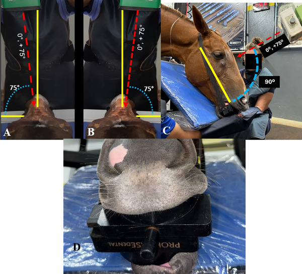
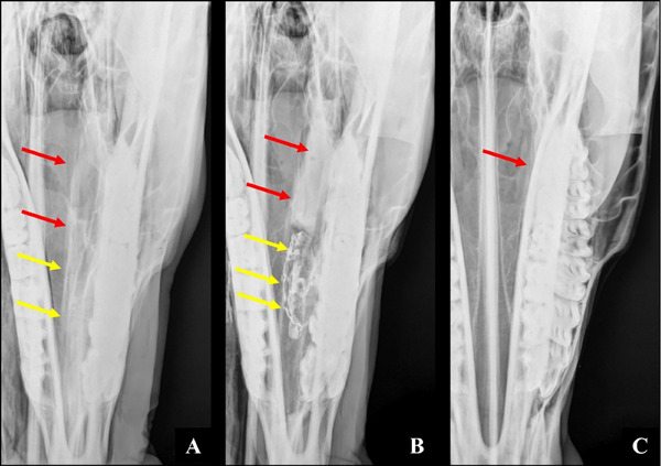
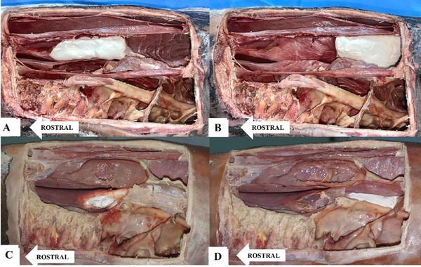
Radiographic examination of the skull is a well-established and indispensable procedure for the diagnostic evaluation of dental and paranasal sinus disorders in horses. Complementary projections make significant contributions to radiographic diagnosis in nasal conchae disorders. This article describes a complementary radiographic projection designed for the evaluation of the conchal sinuses and bullae in horses. Six equine cadaveric heads were used. Specimens were dissected, and openings were created in the ventral and dorsal nasal conchae for the introduction of radiopaque material. The new radiographic projection was named lateral 75° dorsal-lateral ventral oblique view. This projection was obtained with the mandibular rami resting on the imaging plate and the mandible displaced toward the side of interest (partial excursion). The X-ray beam was directed dorsoventrally, slightly angled toward the side of interest (left or right offset mandible dorsoventral view), and centered at an imaginary line connecting the tips of the facial crests. The lateral 75° dorsal-lateral ventral oblique view provided enhanced visualization and allowed correct identification of equine conchal structures. Lateral displacement of the mandible and the 75° angle of inclination to the vertical plane eliminated the superimposition of anatomical structures, facilitating radiographic image interpretation and increasing diagnostic accuracy. This complementary projection is recommended in all cases of sinonasal disorders in horses.
期刊介绍:
Veterinary Radiology & Ultrasound is a bimonthly, international, peer-reviewed, research journal devoted to the fields of veterinary diagnostic imaging and radiation oncology. Established in 1958, it is owned by the American College of Veterinary Radiology and is also the official journal for six affiliate veterinary organizations. Veterinary Radiology & Ultrasound is represented on the International Committee of Medical Journal Editors, World Association of Medical Editors, and Committee on Publication Ethics.
The mission of Veterinary Radiology & Ultrasound is to serve as a leading resource for high quality articles that advance scientific knowledge and standards of clinical practice in the areas of veterinary diagnostic radiology, computed tomography, magnetic resonance imaging, ultrasonography, nuclear imaging, radiation oncology, and interventional radiology. Manuscript types include original investigations, imaging diagnosis reports, review articles, editorials and letters to the Editor. Acceptance criteria include originality, significance, quality, reader interest, composition and adherence to author guidelines.

 求助内容:
求助内容: 应助结果提醒方式:


