{"title":"磷酸化的septin 3从脊柱基部脱位,并通过肌球蛋白- va促进内质网延伸到脊柱。","authors":"Natsumi Ageta-Ishihara, Masato Mizukami, Itsuki Kinoshita, Yurika Asami, Tomoki Nishioka, Haruhiko Bito, Kozo Kaibuchi, Makoto Kinoshita","doi":"10.1186/s13041-025-01215-9","DOIUrl":null,"url":null,"abstract":"<p><p>Cytoskeletal remodeling drives morphological changes. Septin cytoskeleton assembles into hetero-oligomers. We previously demonstrated that late-phase long-term potentiation (L-LTP) induces smooth endoplasmic reticulum (sER) extension into dendritic spines via septin 3 (SEPT3), contributing to greater postsynaptic Ca<sup>2+</sup> responses and enhanced activation of synaptically induced Ca<sup>2+</sup> signaling. Sept3<sup>-/-</sup> mice exhibit a reduced number of sER-containing spines and show impaired long-term spatial/object memory despite normal short-term memory. Additionally, SEPT3 binds the motor protein myosin-Va (MYO5A) upon elevated Ca²⁺ concentrations, facilitating sER extension from the dendritic shaft into the spine. MYO5A localizes on the sER membrane, while SEPT3 remains at the spine base, accumulating on sER upon electroconvulsive stimulation (ECS). However, the mechanism underlying SEPT3's delocalization from the spine base and its cooperative role with MYO5A in sER extension remains unclear. In this study, we demonstrate that SEPT3 is phosphorylated in a stimulation-dependent manner. Phosphorylation at Thr211 releases SEPT3 from the spine base, enabling sER extension with constitutively active MYO5A mutant (MYO5A-CCtr). These findings provide molecular insight into the role of SEPT3 phosphorylation in regulating sER dynamics that sustain long-term spine activation.</p>","PeriodicalId":18851,"journal":{"name":"Molecular Brain","volume":"18 1","pages":"43"},"PeriodicalIF":2.9000,"publicationDate":"2025-05-15","publicationTypes":"Journal Article","fieldsOfStudy":null,"isOpenAccess":false,"openAccessPdf":"https://www.ncbi.nlm.nih.gov/pmc/articles/PMC12079886/pdf/","citationCount":"0","resultStr":"{\"title\":\"Phosphorylated septin 3 delocalizes from the spine base and facilitates endoplasmic reticulum extension into spines via myosin-Va.\",\"authors\":\"Natsumi Ageta-Ishihara, Masato Mizukami, Itsuki Kinoshita, Yurika Asami, Tomoki Nishioka, Haruhiko Bito, Kozo Kaibuchi, Makoto Kinoshita\",\"doi\":\"10.1186/s13041-025-01215-9\",\"DOIUrl\":null,\"url\":null,\"abstract\":\"<p><p>Cytoskeletal remodeling drives morphological changes. Septin cytoskeleton assembles into hetero-oligomers. We previously demonstrated that late-phase long-term potentiation (L-LTP) induces smooth endoplasmic reticulum (sER) extension into dendritic spines via septin 3 (SEPT3), contributing to greater postsynaptic Ca<sup>2+</sup> responses and enhanced activation of synaptically induced Ca<sup>2+</sup> signaling. Sept3<sup>-/-</sup> mice exhibit a reduced number of sER-containing spines and show impaired long-term spatial/object memory despite normal short-term memory. Additionally, SEPT3 binds the motor protein myosin-Va (MYO5A) upon elevated Ca²⁺ concentrations, facilitating sER extension from the dendritic shaft into the spine. MYO5A localizes on the sER membrane, while SEPT3 remains at the spine base, accumulating on sER upon electroconvulsive stimulation (ECS). However, the mechanism underlying SEPT3's delocalization from the spine base and its cooperative role with MYO5A in sER extension remains unclear. In this study, we demonstrate that SEPT3 is phosphorylated in a stimulation-dependent manner. Phosphorylation at Thr211 releases SEPT3 from the spine base, enabling sER extension with constitutively active MYO5A mutant (MYO5A-CCtr). These findings provide molecular insight into the role of SEPT3 phosphorylation in regulating sER dynamics that sustain long-term spine activation.</p>\",\"PeriodicalId\":18851,\"journal\":{\"name\":\"Molecular Brain\",\"volume\":\"18 1\",\"pages\":\"43\"},\"PeriodicalIF\":2.9000,\"publicationDate\":\"2025-05-15\",\"publicationTypes\":\"Journal Article\",\"fieldsOfStudy\":null,\"isOpenAccess\":false,\"openAccessPdf\":\"https://www.ncbi.nlm.nih.gov/pmc/articles/PMC12079886/pdf/\",\"citationCount\":\"0\",\"resultStr\":null,\"platform\":\"Semanticscholar\",\"paperid\":null,\"PeriodicalName\":\"Molecular Brain\",\"FirstCategoryId\":\"3\",\"ListUrlMain\":\"https://doi.org/10.1186/s13041-025-01215-9\",\"RegionNum\":3,\"RegionCategory\":\"医学\",\"ArticlePicture\":[],\"TitleCN\":null,\"AbstractTextCN\":null,\"PMCID\":null,\"EPubDate\":\"\",\"PubModel\":\"\",\"JCR\":\"Q2\",\"JCRName\":\"NEUROSCIENCES\",\"Score\":null,\"Total\":0}","platform":"Semanticscholar","paperid":null,"PeriodicalName":"Molecular Brain","FirstCategoryId":"3","ListUrlMain":"https://doi.org/10.1186/s13041-025-01215-9","RegionNum":3,"RegionCategory":"医学","ArticlePicture":[],"TitleCN":null,"AbstractTextCN":null,"PMCID":null,"EPubDate":"","PubModel":"","JCR":"Q2","JCRName":"NEUROSCIENCES","Score":null,"Total":0}
引用次数: 0
摘要
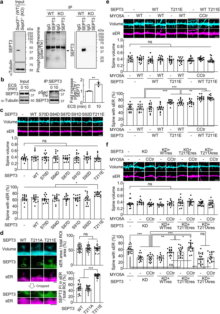
细胞骨架重塑驱动形态改变。Septin细胞骨架组装成异聚物。我们之前已经证明,后期长期增强(L-LTP)通过间隔蛋白3 (SEPT3)诱导光滑内质网(sER)延伸到树突棘,促进了更大的突触后Ca2+反应和突触诱导的Ca2+信号的激活。Sept3-/-小鼠表现出含有ser的脊髓数量减少,尽管短期记忆正常,但长期空间/物体记忆受损。此外,SEPT3在Ca 2 +浓度升高时结合运动蛋白肌球蛋白- va (MYO5A),促进sER从树突轴延伸到脊柱。MYO5A位于sER膜上,而SEPT3则位于脊柱基部,在电痉挛刺激(ECS)时积聚在sER上。然而,SEPT3从脊柱基部脱位的机制及其与MYO5A在sER扩展中的合作作用尚不清楚。在这项研究中,我们证明了SEPT3以刺激依赖的方式磷酸化。Thr211位点的磷酸化释放了脊柱基部的SEPT3,使得具有组成性活性MYO5A突变体(MYO5A- cctr)的sER延伸成为可能。这些发现为SEPT3磷酸化在调节维持长期脊柱激活的sER动力学中的作用提供了分子视角。
Phosphorylated septin 3 delocalizes from the spine base and facilitates endoplasmic reticulum extension into spines via myosin-Va.
Cytoskeletal remodeling drives morphological changes. Septin cytoskeleton assembles into hetero-oligomers. We previously demonstrated that late-phase long-term potentiation (L-LTP) induces smooth endoplasmic reticulum (sER) extension into dendritic spines via septin 3 (SEPT3), contributing to greater postsynaptic Ca2+ responses and enhanced activation of synaptically induced Ca2+ signaling. Sept3-/- mice exhibit a reduced number of sER-containing spines and show impaired long-term spatial/object memory despite normal short-term memory. Additionally, SEPT3 binds the motor protein myosin-Va (MYO5A) upon elevated Ca²⁺ concentrations, facilitating sER extension from the dendritic shaft into the spine. MYO5A localizes on the sER membrane, while SEPT3 remains at the spine base, accumulating on sER upon electroconvulsive stimulation (ECS). However, the mechanism underlying SEPT3's delocalization from the spine base and its cooperative role with MYO5A in sER extension remains unclear. In this study, we demonstrate that SEPT3 is phosphorylated in a stimulation-dependent manner. Phosphorylation at Thr211 releases SEPT3 from the spine base, enabling sER extension with constitutively active MYO5A mutant (MYO5A-CCtr). These findings provide molecular insight into the role of SEPT3 phosphorylation in regulating sER dynamics that sustain long-term spine activation.
期刊介绍:
Molecular Brain is an open access, peer-reviewed journal that considers manuscripts on all aspects of studies on the nervous system at the molecular, cellular, and systems level providing a forum for scientists to communicate their findings.
Molecular brain research is a rapidly expanding research field in which integrative approaches at the genetic, molecular, cellular and synaptic levels yield key information about the physiological and pathological brain. These studies involve the use of a wide range of modern techniques in molecular biology, genomics, proteomics, imaging and electrophysiology.
 求助内容:
求助内容: 应助结果提醒方式:


