{"title":"视网膜外周孔引起的特发性和继发性视网膜上膜的光学相干断层扫描特征。","authors":"Yuanyuan Fan, Yingying Jiang, Zhaoxia Mu, Yulian Xu, Ping Xie, Qinghuai Liu, Lijun Pu, Zizhong Hu","doi":"10.1155/joph/9299651","DOIUrl":null,"url":null,"abstract":"<p><p><b>Purpose:</b> In clinical practice, some eyes preoperatively diagnosed with \"idiopathic epiretinal membranes (iERM)\" will be amended to \"secondary epiretinal membranes (sERM)\" once peripheral retinal hole is detected. This study utilized optical coherence tomography (OCT) images to compare the characteristics between the iERM and sERM due to peripheral retinal hole (PRH). <b>Methods:</b> In this retrospective, cross-sectional study, 635 eyes that had undergone pars plana vitrectomy with membrane peeling were enrolled. A total of 115 eyes (18.1%) detected with peripheral retinal holes were allocated to the sERM-PRH group while the other 520 eyes were to the iERM group. The demographic data and OCT characteristics were compared between the two groups. Besides, all the eyes were evaluated by a double-grading scheme: severity grading of ERM progression into four stages plus anatomical classification into three kinds of part-thickness macular holes associated with ERMs. <b>Results:</b> No significant difference was found in age, gender, symptom duration, axial length, or best-corrected visual acuity between the two groups. There was also no difference concerning the features based on OCT, ranging from central macular thickness, the ratios of the photoreceptor inner/outer segment junction line defect, intraretinal fluid, cotton ball sign, to epiretinal proliferation. However, the native difference in parafoveal thickness between the temporal and nasal quadrants was observed in the iERM group, yet disappeared in the sERM-PRH group. Moreover, eyes between the two groups were distributionally similar in both grading scales. <b>Conclusion:</b> Our results demonstrated that even OCT images could hardly provide effective clues for early differentiating sERM from iERM, which highlighted the necessity of a thorough pre- and intro-operative fundus examination of the peripheral retina for clinicians.</p>","PeriodicalId":16674,"journal":{"name":"Journal of Ophthalmology","volume":"2025 ","pages":"9299651"},"PeriodicalIF":1.9000,"publicationDate":"2025-05-07","publicationTypes":"Journal Article","fieldsOfStudy":null,"isOpenAccess":false,"openAccessPdf":"https://www.ncbi.nlm.nih.gov/pmc/articles/PMC12077978/pdf/","citationCount":"0","resultStr":"{\"title\":\"Optical Coherence Tomography Characteristics Between Idiopathic Epiretinal Membranes and Secondary Epiretinal Membranes due to Peripheral Retinal Hole.\",\"authors\":\"Yuanyuan Fan, Yingying Jiang, Zhaoxia Mu, Yulian Xu, Ping Xie, Qinghuai Liu, Lijun Pu, Zizhong Hu\",\"doi\":\"10.1155/joph/9299651\",\"DOIUrl\":null,\"url\":null,\"abstract\":\"<p><p><b>Purpose:</b> In clinical practice, some eyes preoperatively diagnosed with \\\"idiopathic epiretinal membranes (iERM)\\\" will be amended to \\\"secondary epiretinal membranes (sERM)\\\" once peripheral retinal hole is detected. This study utilized optical coherence tomography (OCT) images to compare the characteristics between the iERM and sERM due to peripheral retinal hole (PRH). <b>Methods:</b> In this retrospective, cross-sectional study, 635 eyes that had undergone pars plana vitrectomy with membrane peeling were enrolled. A total of 115 eyes (18.1%) detected with peripheral retinal holes were allocated to the sERM-PRH group while the other 520 eyes were to the iERM group. The demographic data and OCT characteristics were compared between the two groups. Besides, all the eyes were evaluated by a double-grading scheme: severity grading of ERM progression into four stages plus anatomical classification into three kinds of part-thickness macular holes associated with ERMs. <b>Results:</b> No significant difference was found in age, gender, symptom duration, axial length, or best-corrected visual acuity between the two groups. There was also no difference concerning the features based on OCT, ranging from central macular thickness, the ratios of the photoreceptor inner/outer segment junction line defect, intraretinal fluid, cotton ball sign, to epiretinal proliferation. However, the native difference in parafoveal thickness between the temporal and nasal quadrants was observed in the iERM group, yet disappeared in the sERM-PRH group. Moreover, eyes between the two groups were distributionally similar in both grading scales. <b>Conclusion:</b> Our results demonstrated that even OCT images could hardly provide effective clues for early differentiating sERM from iERM, which highlighted the necessity of a thorough pre- and intro-operative fundus examination of the peripheral retina for clinicians.</p>\",\"PeriodicalId\":16674,\"journal\":{\"name\":\"Journal of Ophthalmology\",\"volume\":\"2025 \",\"pages\":\"9299651\"},\"PeriodicalIF\":1.9000,\"publicationDate\":\"2025-05-07\",\"publicationTypes\":\"Journal Article\",\"fieldsOfStudy\":null,\"isOpenAccess\":false,\"openAccessPdf\":\"https://www.ncbi.nlm.nih.gov/pmc/articles/PMC12077978/pdf/\",\"citationCount\":\"0\",\"resultStr\":null,\"platform\":\"Semanticscholar\",\"paperid\":null,\"PeriodicalName\":\"Journal of Ophthalmology\",\"FirstCategoryId\":\"3\",\"ListUrlMain\":\"https://doi.org/10.1155/joph/9299651\",\"RegionNum\":4,\"RegionCategory\":\"医学\",\"ArticlePicture\":[],\"TitleCN\":null,\"AbstractTextCN\":null,\"PMCID\":null,\"EPubDate\":\"2025/1/1 0:00:00\",\"PubModel\":\"eCollection\",\"JCR\":\"Q3\",\"JCRName\":\"OPHTHALMOLOGY\",\"Score\":null,\"Total\":0}","platform":"Semanticscholar","paperid":null,"PeriodicalName":"Journal of Ophthalmology","FirstCategoryId":"3","ListUrlMain":"https://doi.org/10.1155/joph/9299651","RegionNum":4,"RegionCategory":"医学","ArticlePicture":[],"TitleCN":null,"AbstractTextCN":null,"PMCID":null,"EPubDate":"2025/1/1 0:00:00","PubModel":"eCollection","JCR":"Q3","JCRName":"OPHTHALMOLOGY","Score":null,"Total":0}
Optical Coherence Tomography Characteristics Between Idiopathic Epiretinal Membranes and Secondary Epiretinal Membranes due to Peripheral Retinal Hole.
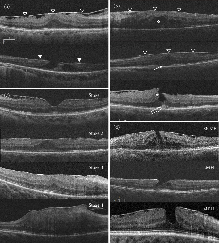
Purpose: In clinical practice, some eyes preoperatively diagnosed with "idiopathic epiretinal membranes (iERM)" will be amended to "secondary epiretinal membranes (sERM)" once peripheral retinal hole is detected. This study utilized optical coherence tomography (OCT) images to compare the characteristics between the iERM and sERM due to peripheral retinal hole (PRH). Methods: In this retrospective, cross-sectional study, 635 eyes that had undergone pars plana vitrectomy with membrane peeling were enrolled. A total of 115 eyes (18.1%) detected with peripheral retinal holes were allocated to the sERM-PRH group while the other 520 eyes were to the iERM group. The demographic data and OCT characteristics were compared between the two groups. Besides, all the eyes were evaluated by a double-grading scheme: severity grading of ERM progression into four stages plus anatomical classification into three kinds of part-thickness macular holes associated with ERMs. Results: No significant difference was found in age, gender, symptom duration, axial length, or best-corrected visual acuity between the two groups. There was also no difference concerning the features based on OCT, ranging from central macular thickness, the ratios of the photoreceptor inner/outer segment junction line defect, intraretinal fluid, cotton ball sign, to epiretinal proliferation. However, the native difference in parafoveal thickness between the temporal and nasal quadrants was observed in the iERM group, yet disappeared in the sERM-PRH group. Moreover, eyes between the two groups were distributionally similar in both grading scales. Conclusion: Our results demonstrated that even OCT images could hardly provide effective clues for early differentiating sERM from iERM, which highlighted the necessity of a thorough pre- and intro-operative fundus examination of the peripheral retina for clinicians.
期刊介绍:
Journal of Ophthalmology is a peer-reviewed, Open Access journal that publishes original research articles, review articles, and clinical studies related to the anatomy, physiology and diseases of the eye. Submissions should focus on new diagnostic and surgical techniques, instrument and therapy updates, as well as clinical trials and research findings.
 求助内容:
求助内容: 应助结果提醒方式:


