Yi Sun, Yuanyuan Chen, Jing Zhu, Juan Guo, Zhanfeng Wang
{"title":"虹膜睫状体黑色素细胞瘤伴继发性青光眼1例。","authors":"Yi Sun, Yuanyuan Chen, Jing Zhu, Juan Guo, Zhanfeng Wang","doi":"10.1186/s13000-025-01646-x","DOIUrl":null,"url":null,"abstract":"<p><strong>Background: </strong>Distinguishing between benign iridociliary melanocytoma and malignant melanoma presents a diagnostic challenge, particularly given the potential overlap in tumor growth patterns and clinical manifestations, especially when patients present with secondary glaucoma. Misdiagnosis may induce severe clinical consequences, including enucleation. Therefore, the judicious selection of biopsy or surgical techniques is crucial in both diagnosing and managing the condition.</p><p><strong>Case presentation: </strong>A 44-year-old female presented with uncontrolled elevated intraocular pressure (IOP) and a heavily pigmented iris lesion extending into the anterior chamber angle and adjacent ciliary body. Unexpectedly, standardized initial fine-needle aspiration biopsy (FNAB) yielded inconclusive results. Subsequent excisional surgery (partial iridocyclectomy and concurrent phacoemulsification) was performed to remove the tumor mass and treat cataract. Histopathological analysis confirmed the diagnosis as melanocytoma. Lens implantation followed upon normalization of IOP within 8 months. At the 2-year follow-up, the patient exhibited a satisfactory clinical outcome, with no tumor recurrence, achieving a best-corrected visual acuity of 20/40 and an intraocular pressure of 18.5 mmHg.</p><p><strong>Conclusions: </strong>This case underscores the importance of obtaining adequate tumor specimens for accurate diagnosis via FNAB in iris and ciliary body tumors. Additionally, for patients with secondary glaucoma, partial iridocyclectomy emerges as a promising intervention, addressing anterior chamber angle obstruction to alleviate IOP while facilitating histopathological diagnosis for subsequent management.</p>","PeriodicalId":11237,"journal":{"name":"Diagnostic Pathology","volume":"20 1","pages":"60"},"PeriodicalIF":2.3000,"publicationDate":"2025-05-15","publicationTypes":"Journal Article","fieldsOfStudy":null,"isOpenAccess":false,"openAccessPdf":"https://www.ncbi.nlm.nih.gov/pmc/articles/PMC12082992/pdf/","citationCount":"0","resultStr":"{\"title\":\"A case report: iridociliary melanocytoma associated with secondary glaucoma.\",\"authors\":\"Yi Sun, Yuanyuan Chen, Jing Zhu, Juan Guo, Zhanfeng Wang\",\"doi\":\"10.1186/s13000-025-01646-x\",\"DOIUrl\":null,\"url\":null,\"abstract\":\"<p><strong>Background: </strong>Distinguishing between benign iridociliary melanocytoma and malignant melanoma presents a diagnostic challenge, particularly given the potential overlap in tumor growth patterns and clinical manifestations, especially when patients present with secondary glaucoma. Misdiagnosis may induce severe clinical consequences, including enucleation. Therefore, the judicious selection of biopsy or surgical techniques is crucial in both diagnosing and managing the condition.</p><p><strong>Case presentation: </strong>A 44-year-old female presented with uncontrolled elevated intraocular pressure (IOP) and a heavily pigmented iris lesion extending into the anterior chamber angle and adjacent ciliary body. Unexpectedly, standardized initial fine-needle aspiration biopsy (FNAB) yielded inconclusive results. Subsequent excisional surgery (partial iridocyclectomy and concurrent phacoemulsification) was performed to remove the tumor mass and treat cataract. Histopathological analysis confirmed the diagnosis as melanocytoma. Lens implantation followed upon normalization of IOP within 8 months. At the 2-year follow-up, the patient exhibited a satisfactory clinical outcome, with no tumor recurrence, achieving a best-corrected visual acuity of 20/40 and an intraocular pressure of 18.5 mmHg.</p><p><strong>Conclusions: </strong>This case underscores the importance of obtaining adequate tumor specimens for accurate diagnosis via FNAB in iris and ciliary body tumors. Additionally, for patients with secondary glaucoma, partial iridocyclectomy emerges as a promising intervention, addressing anterior chamber angle obstruction to alleviate IOP while facilitating histopathological diagnosis for subsequent management.</p>\",\"PeriodicalId\":11237,\"journal\":{\"name\":\"Diagnostic Pathology\",\"volume\":\"20 1\",\"pages\":\"60\"},\"PeriodicalIF\":2.3000,\"publicationDate\":\"2025-05-15\",\"publicationTypes\":\"Journal Article\",\"fieldsOfStudy\":null,\"isOpenAccess\":false,\"openAccessPdf\":\"https://www.ncbi.nlm.nih.gov/pmc/articles/PMC12082992/pdf/\",\"citationCount\":\"0\",\"resultStr\":null,\"platform\":\"Semanticscholar\",\"paperid\":null,\"PeriodicalName\":\"Diagnostic Pathology\",\"FirstCategoryId\":\"3\",\"ListUrlMain\":\"https://doi.org/10.1186/s13000-025-01646-x\",\"RegionNum\":3,\"RegionCategory\":\"医学\",\"ArticlePicture\":[],\"TitleCN\":null,\"AbstractTextCN\":null,\"PMCID\":null,\"EPubDate\":\"\",\"PubModel\":\"\",\"JCR\":\"Q2\",\"JCRName\":\"PATHOLOGY\",\"Score\":null,\"Total\":0}","platform":"Semanticscholar","paperid":null,"PeriodicalName":"Diagnostic Pathology","FirstCategoryId":"3","ListUrlMain":"https://doi.org/10.1186/s13000-025-01646-x","RegionNum":3,"RegionCategory":"医学","ArticlePicture":[],"TitleCN":null,"AbstractTextCN":null,"PMCID":null,"EPubDate":"","PubModel":"","JCR":"Q2","JCRName":"PATHOLOGY","Score":null,"Total":0}
A case report: iridociliary melanocytoma associated with secondary glaucoma.
Background: Distinguishing between benign iridociliary melanocytoma and malignant melanoma presents a diagnostic challenge, particularly given the potential overlap in tumor growth patterns and clinical manifestations, especially when patients present with secondary glaucoma. Misdiagnosis may induce severe clinical consequences, including enucleation. Therefore, the judicious selection of biopsy or surgical techniques is crucial in both diagnosing and managing the condition.
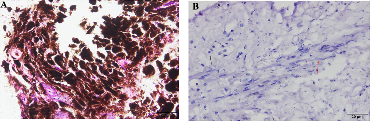
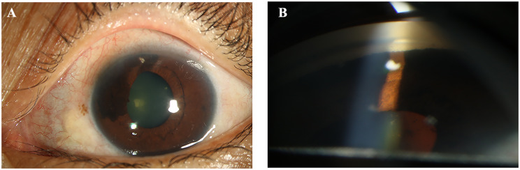
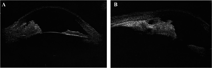
Case presentation: A 44-year-old female presented with uncontrolled elevated intraocular pressure (IOP) and a heavily pigmented iris lesion extending into the anterior chamber angle and adjacent ciliary body. Unexpectedly, standardized initial fine-needle aspiration biopsy (FNAB) yielded inconclusive results. Subsequent excisional surgery (partial iridocyclectomy and concurrent phacoemulsification) was performed to remove the tumor mass and treat cataract. Histopathological analysis confirmed the diagnosis as melanocytoma. Lens implantation followed upon normalization of IOP within 8 months. At the 2-year follow-up, the patient exhibited a satisfactory clinical outcome, with no tumor recurrence, achieving a best-corrected visual acuity of 20/40 and an intraocular pressure of 18.5 mmHg.
Conclusions: This case underscores the importance of obtaining adequate tumor specimens for accurate diagnosis via FNAB in iris and ciliary body tumors. Additionally, for patients with secondary glaucoma, partial iridocyclectomy emerges as a promising intervention, addressing anterior chamber angle obstruction to alleviate IOP while facilitating histopathological diagnosis for subsequent management.
期刊介绍:
Diagnostic Pathology is an open access, peer-reviewed, online journal that considers research in surgical and clinical pathology, immunology, and biology, with a special focus on cutting-edge approaches in diagnostic pathology and tissue-based therapy. The journal covers all aspects of surgical pathology, including classic diagnostic pathology, prognosis-related diagnosis (tumor stages, prognosis markers, such as MIB-percentage, hormone receptors, etc.), and therapy-related findings. The journal also focuses on the technological aspects of pathology, including molecular biology techniques, morphometry aspects (stereology, DNA analysis, syntactic structure analysis), communication aspects (telecommunication, virtual microscopy, virtual pathology institutions, etc.), and electronic education and quality assurance (for example interactive publication, on-line references with automated updating, etc.).

 求助内容:
求助内容: 应助结果提醒方式:


