Zita Matias, Catarina S Lopes, Nuno C Santos, Filomena A Carvalho
{"title":"纳米技术与医学:原子力显微镜在疾病中的应用。","authors":"Zita Matias, Catarina S Lopes, Nuno C Santos, Filomena A Carvalho","doi":"10.1007/s12551-025-01306-w","DOIUrl":null,"url":null,"abstract":"<p><p>Atomic force microscopy (AFM) is a scanning imaging technique able to work at the nanoscale. It uses a cantilever with a tip to move across samples' surface and a laser to measure the cantilever bending, enabling the assessment of interaction forces between tip and sample and creating a three-dimensional visual representation of its surface. AFM has been gaining notoriety in the biomedical field due to its high-resolution images, as well as due to its ability to measure the inter- and intramolecular interaction forces involved in the pathophysiology of many diseases. Here, we highlight some of the current applications of AFM in the biomedical field. First, a brief overview of the AFM technique is presented. This theoretical framework is then used to link AFM to its novel translational applications, handling broad clinical questions in different areas, such as infectious diseases, cardiovascular diseases, cancer, and neurodegenerative diseases. Morphological and nanomechanical characteristics such as cell height, volume, stiffness, and adhesion forces may serve as novel parameters used to tailor patient care through nanodiagnostics, individualized risk stratification, and therapeutic monitoring. Despite an increasing development of AFM biomedical research with patient cells, showing its unique capabilities in terms of resolution, speed, and accuracy, there is a notable need for applied AFM research in clinical settings. More translational research with AFM may provide new grounds for the valuable collaboration between biomedical researchers and healthcare professionals.</p>","PeriodicalId":9094,"journal":{"name":"Biophysical reviews","volume":"17 2","pages":"359-384"},"PeriodicalIF":3.7000,"publicationDate":"2025-04-03","publicationTypes":"Journal Article","fieldsOfStudy":null,"isOpenAccess":false,"openAccessPdf":"https://www.ncbi.nlm.nih.gov/pmc/articles/PMC12075069/pdf/","citationCount":"0","resultStr":"{\"title\":\"Nanotechnology meets medicine: applications of atomic force microscopy in disease.\",\"authors\":\"Zita Matias, Catarina S Lopes, Nuno C Santos, Filomena A Carvalho\",\"doi\":\"10.1007/s12551-025-01306-w\",\"DOIUrl\":null,\"url\":null,\"abstract\":\"<p><p>Atomic force microscopy (AFM) is a scanning imaging technique able to work at the nanoscale. It uses a cantilever with a tip to move across samples' surface and a laser to measure the cantilever bending, enabling the assessment of interaction forces between tip and sample and creating a three-dimensional visual representation of its surface. AFM has been gaining notoriety in the biomedical field due to its high-resolution images, as well as due to its ability to measure the inter- and intramolecular interaction forces involved in the pathophysiology of many diseases. Here, we highlight some of the current applications of AFM in the biomedical field. First, a brief overview of the AFM technique is presented. This theoretical framework is then used to link AFM to its novel translational applications, handling broad clinical questions in different areas, such as infectious diseases, cardiovascular diseases, cancer, and neurodegenerative diseases. Morphological and nanomechanical characteristics such as cell height, volume, stiffness, and adhesion forces may serve as novel parameters used to tailor patient care through nanodiagnostics, individualized risk stratification, and therapeutic monitoring. Despite an increasing development of AFM biomedical research with patient cells, showing its unique capabilities in terms of resolution, speed, and accuracy, there is a notable need for applied AFM research in clinical settings. More translational research with AFM may provide new grounds for the valuable collaboration between biomedical researchers and healthcare professionals.</p>\",\"PeriodicalId\":9094,\"journal\":{\"name\":\"Biophysical reviews\",\"volume\":\"17 2\",\"pages\":\"359-384\"},\"PeriodicalIF\":3.7000,\"publicationDate\":\"2025-04-03\",\"publicationTypes\":\"Journal Article\",\"fieldsOfStudy\":null,\"isOpenAccess\":false,\"openAccessPdf\":\"https://www.ncbi.nlm.nih.gov/pmc/articles/PMC12075069/pdf/\",\"citationCount\":\"0\",\"resultStr\":null,\"platform\":\"Semanticscholar\",\"paperid\":null,\"PeriodicalName\":\"Biophysical reviews\",\"FirstCategoryId\":\"1085\",\"ListUrlMain\":\"https://doi.org/10.1007/s12551-025-01306-w\",\"RegionNum\":0,\"RegionCategory\":null,\"ArticlePicture\":[],\"TitleCN\":null,\"AbstractTextCN\":null,\"PMCID\":null,\"EPubDate\":\"2025/4/1 0:00:00\",\"PubModel\":\"eCollection\",\"JCR\":\"Q1\",\"JCRName\":\"BIOPHYSICS\",\"Score\":null,\"Total\":0}","platform":"Semanticscholar","paperid":null,"PeriodicalName":"Biophysical reviews","FirstCategoryId":"1085","ListUrlMain":"https://doi.org/10.1007/s12551-025-01306-w","RegionNum":0,"RegionCategory":null,"ArticlePicture":[],"TitleCN":null,"AbstractTextCN":null,"PMCID":null,"EPubDate":"2025/4/1 0:00:00","PubModel":"eCollection","JCR":"Q1","JCRName":"BIOPHYSICS","Score":null,"Total":0}
Nanotechnology meets medicine: applications of atomic force microscopy in disease.
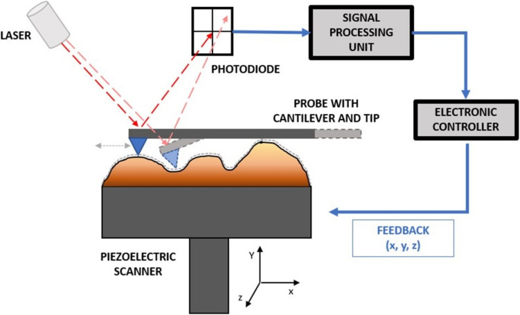
Atomic force microscopy (AFM) is a scanning imaging technique able to work at the nanoscale. It uses a cantilever with a tip to move across samples' surface and a laser to measure the cantilever bending, enabling the assessment of interaction forces between tip and sample and creating a three-dimensional visual representation of its surface. AFM has been gaining notoriety in the biomedical field due to its high-resolution images, as well as due to its ability to measure the inter- and intramolecular interaction forces involved in the pathophysiology of many diseases. Here, we highlight some of the current applications of AFM in the biomedical field. First, a brief overview of the AFM technique is presented. This theoretical framework is then used to link AFM to its novel translational applications, handling broad clinical questions in different areas, such as infectious diseases, cardiovascular diseases, cancer, and neurodegenerative diseases. Morphological and nanomechanical characteristics such as cell height, volume, stiffness, and adhesion forces may serve as novel parameters used to tailor patient care through nanodiagnostics, individualized risk stratification, and therapeutic monitoring. Despite an increasing development of AFM biomedical research with patient cells, showing its unique capabilities in terms of resolution, speed, and accuracy, there is a notable need for applied AFM research in clinical settings. More translational research with AFM may provide new grounds for the valuable collaboration between biomedical researchers and healthcare professionals.
期刊介绍:
Biophysical Reviews aims to publish critical and timely reviews from key figures in the field of biophysics. The bulk of the reviews that are currently published are from invited authors, but the journal is also open for non-solicited reviews. Interested authors are encouraged to discuss the possibility of contributing a review with the Editor-in-Chief prior to submission. Through publishing reviews on biophysics, the editors of the journal hope to illustrate the great power and potential of physical techniques in the biological sciences, they aim to stimulate the discussion and promote further research and would like to educate and enthuse basic researcher scientists and students of biophysics. Biophysical Reviews covers the entire field of biophysics, generally defined as the science of describing and defining biological phenomenon using the concepts and the techniques of physics. This includes but is not limited by such areas as: - Bioinformatics - Biophysical methods and instrumentation - Medical biophysics - Biosystems - Cell biophysics and organization - Macromolecules: dynamics, structures and interactions - Single molecule biophysics - Membrane biophysics, channels and transportation

 求助内容:
求助内容: 应助结果提醒方式:


