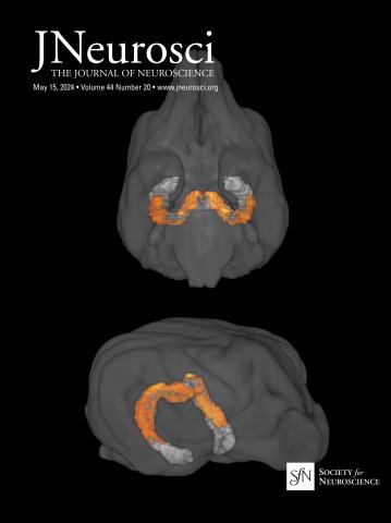
猴鼻周围神经元投射到尾状核的化学发生破坏损害了联想学习。
IF 4.4
2区 医学
Q1 NEUROSCIENCES
引用次数: 0
摘要
灵长类动物,包括人类,使用刺激-奖励关联来指导觅食。我们以前的研究表明,恒河猴的鼻皮质(Rh)和rostromedial尾状核(rmCD)在视觉刺激的价值分配中起因果作用。Rh第5层神经元向rmCD投射。在这里,我们通过将逆行慢病毒(FuG-E)注射到rmCD中,将单侧Rh病变与对侧抑制性DREADD表达结合,可逆地中断了两只雄性猴子的第5层连接。从Rh到rmCD的投射神经元中断对已经学习的刺激-奖励关联影响不大,但会损害新关联的学习。将去氯氯氮平(DCZ)微量注射到外周皮层(PRh)后,使外周皮质(PRh)向rmCD的投射神经元沉默,出现学习障碍。通路特异性沉默导致学习新的刺激-奖励关联的显著缺陷。这些结果表明,视觉刺激-奖励关联的学习,而不是检索,涉及从PRh到rmCD的投射神经元。灵长类动物使用刺激-奖励关联来指导觅食。Rh和rmCD在非人类灵长类动物的视觉刺激-奖励关联中都起着因果作用。从Rh到rmCD有很强的解剖投影。通过向rmCD中注射逆行慢病毒(FuG-E),将单侧Rh病变与对侧抑制性DREADD表达结合起来,这种联系被可逆地中断。这种联系的中断对已经习得的刺激-奖励联系几乎没有影响,但会损害新联系的学习。我们进一步将这种学习障碍的来源定位为从Rh(即PRh)投射到rmCD的神经元。我们的研究结果强调了该回路在适应性行为中的重要性,并表明学习和检索依赖于不同的神经通路。本文章由计算机程序翻译,如有差异,请以英文原文为准。
Chemogenetic disruption of monkey perirhinal neurons projecting to rostromedial caudate impairs associative learning.
Primates, including humans, use stimulus-reward associations to guide foraging. We previously showed that both rhinal cortex (Rh) and rostromedial caudate (rmCD) of rhesus monkeys play causal roles in assigning value to visual stimuli. Layer 5 neurons in Rh project to rmCD. Here, we reversibly interrupted this layer 5 connection in two male monkeys by combining a unilateral Rh lesion with contralateral expression of an inhibitory DREADD delivered using a retrograde lentivirus (FuG-E) injected into rmCD. Interruption of projection neurons from Rh to rmCD had little effect on already learned stimulus-reward associations but impaired the learning of new associations. The learning impairment appeared when the projection neurons from perirhinal cortex (PRh) to rmCD were silenced using microinjections of deschloroclozapine (DCZ) into PRh. The pathway-specific silencing led to a significant deficit in learning new stimulus-reward associations. These results suggest that the learning, but not retrieval, of visual stimulus-reward associations involves projection neurons from PRh to rmCD.Significance Statement Primates use stimulus-reward associations to guide foraging. Both Rh and rmCD play a causal role in visual-stimulus-reward associations in non-human primates. There is a strong anatomical projection from Rh to rmCD. This connection was reversibly interrupted by combining a unilateral Rh lesion with contralateral expression of an inhibitory DREADD via a retrograde lentivirus (FuG-E) injected into rmCD. Interruption of this connection had little effect on already learned stimulus-reward associations but impaired the learning of new associations. We further localized the source of this learning impairment to neurons projecting to rmCD from the Rh, i.e., PRh. Our findings emphasize the significance of this circuit in adaptive behavior and indicate that learning and retrieval depend on different neural pathways.
求助全文
通过发布文献求助,成功后即可免费获取论文全文。
去求助
来源期刊
Journal of Neuroscience
医学-神经科学
CiteScore
9.30
自引率
3.80%
发文量
1164
审稿时长
12 months
期刊介绍:
JNeurosci (ISSN 0270-6474) is an official journal of the Society for Neuroscience. It is published weekly by the Society, fifty weeks a year, one volume a year. JNeurosci publishes papers on a broad range of topics of general interest to those working on the nervous system. Authors now have an Open Choice option for their published articles
 求助内容:
求助内容: 应助结果提醒方式:


