David Andrew Prentice, Ravi Ambati, Lay K Kho, Thomas Jenkins, Paul M Parizel
{"title":"血清阴性自身免疫性脑脊髓炎伴区域后发症状。","authors":"David Andrew Prentice, Ravi Ambati, Lay K Kho, Thomas Jenkins, Paul M Parizel","doi":"10.1159/000545402","DOIUrl":null,"url":null,"abstract":"<p><strong>Introduction: </strong>Patients presenting with encephalopathy and longitudinally extensive myelitis pose a significant diagnostic challenge. Area postrema-related symptoms, such as intractable hiccoughs, can aid in narrowing the differential diagnosis. Neuromyelitis optica spectrum disorders and glial fibrillary acidic protein (GFAP) autoimmune encephalitis are known causes; however, some cases remain seronegative, suggesting the presence of unidentified autoantibodies or immune targets.</p><p><strong>Case presentation: </strong>A previously healthy man in his 70s presented with headache, fever, and confusion, followed by a seizure and persistent hiccoughs. MRI revealed brainstem involvement and extensive transverse myelitis. Cerebrospinal fluid (CSF) analysis showed inflammatory features, but testing for AQP4, MOG, and GFAP antibodies was initially negative. He was treated with intravenous corticosteroids and plasma exchange, after which serum GFAP-IgG was weakly positive, though CSF remained negative. His condition improved with immunotherapy, but significant lower limb weakness persisted. Based on clinical and radiological findings, we hypothesize that tanycytes - specialized glial cells in the area postrema - may be an additional immune target in GFAP encephalitis.</p><p><strong>Conclusion: </strong>This case highlights a seronegative encephalomyelitis syndrome with area postrema involvement, possibly implicating glial cells beyond astrocytes. Further studies are needed to explore the role of tanycytes in autoimmune neuroinflammation.</p>","PeriodicalId":9639,"journal":{"name":"Case Reports in Neurology","volume":"17 1","pages":"50-56"},"PeriodicalIF":0.6000,"publicationDate":"2025-03-26","publicationTypes":"Journal Article","fieldsOfStudy":null,"isOpenAccess":false,"openAccessPdf":"https://www.ncbi.nlm.nih.gov/pmc/articles/PMC12058113/pdf/","citationCount":"0","resultStr":"{\"title\":\"Seronegative Autoimmune Encephalomyelitis with Area Postrema Symptoms.\",\"authors\":\"David Andrew Prentice, Ravi Ambati, Lay K Kho, Thomas Jenkins, Paul M Parizel\",\"doi\":\"10.1159/000545402\",\"DOIUrl\":null,\"url\":null,\"abstract\":\"<p><strong>Introduction: </strong>Patients presenting with encephalopathy and longitudinally extensive myelitis pose a significant diagnostic challenge. Area postrema-related symptoms, such as intractable hiccoughs, can aid in narrowing the differential diagnosis. Neuromyelitis optica spectrum disorders and glial fibrillary acidic protein (GFAP) autoimmune encephalitis are known causes; however, some cases remain seronegative, suggesting the presence of unidentified autoantibodies or immune targets.</p><p><strong>Case presentation: </strong>A previously healthy man in his 70s presented with headache, fever, and confusion, followed by a seizure and persistent hiccoughs. MRI revealed brainstem involvement and extensive transverse myelitis. Cerebrospinal fluid (CSF) analysis showed inflammatory features, but testing for AQP4, MOG, and GFAP antibodies was initially negative. He was treated with intravenous corticosteroids and plasma exchange, after which serum GFAP-IgG was weakly positive, though CSF remained negative. His condition improved with immunotherapy, but significant lower limb weakness persisted. Based on clinical and radiological findings, we hypothesize that tanycytes - specialized glial cells in the area postrema - may be an additional immune target in GFAP encephalitis.</p><p><strong>Conclusion: </strong>This case highlights a seronegative encephalomyelitis syndrome with area postrema involvement, possibly implicating glial cells beyond astrocytes. Further studies are needed to explore the role of tanycytes in autoimmune neuroinflammation.</p>\",\"PeriodicalId\":9639,\"journal\":{\"name\":\"Case Reports in Neurology\",\"volume\":\"17 1\",\"pages\":\"50-56\"},\"PeriodicalIF\":0.6000,\"publicationDate\":\"2025-03-26\",\"publicationTypes\":\"Journal Article\",\"fieldsOfStudy\":null,\"isOpenAccess\":false,\"openAccessPdf\":\"https://www.ncbi.nlm.nih.gov/pmc/articles/PMC12058113/pdf/\",\"citationCount\":\"0\",\"resultStr\":null,\"platform\":\"Semanticscholar\",\"paperid\":null,\"PeriodicalName\":\"Case Reports in Neurology\",\"FirstCategoryId\":\"1085\",\"ListUrlMain\":\"https://doi.org/10.1159/000545402\",\"RegionNum\":0,\"RegionCategory\":null,\"ArticlePicture\":[],\"TitleCN\":null,\"AbstractTextCN\":null,\"PMCID\":null,\"EPubDate\":\"2025/1/1 0:00:00\",\"PubModel\":\"eCollection\",\"JCR\":\"Q4\",\"JCRName\":\"CLINICAL NEUROLOGY\",\"Score\":null,\"Total\":0}","platform":"Semanticscholar","paperid":null,"PeriodicalName":"Case Reports in Neurology","FirstCategoryId":"1085","ListUrlMain":"https://doi.org/10.1159/000545402","RegionNum":0,"RegionCategory":null,"ArticlePicture":[],"TitleCN":null,"AbstractTextCN":null,"PMCID":null,"EPubDate":"2025/1/1 0:00:00","PubModel":"eCollection","JCR":"Q4","JCRName":"CLINICAL NEUROLOGY","Score":null,"Total":0}
Seronegative Autoimmune Encephalomyelitis with Area Postrema Symptoms.
Introduction: Patients presenting with encephalopathy and longitudinally extensive myelitis pose a significant diagnostic challenge. Area postrema-related symptoms, such as intractable hiccoughs, can aid in narrowing the differential diagnosis. Neuromyelitis optica spectrum disorders and glial fibrillary acidic protein (GFAP) autoimmune encephalitis are known causes; however, some cases remain seronegative, suggesting the presence of unidentified autoantibodies or immune targets.
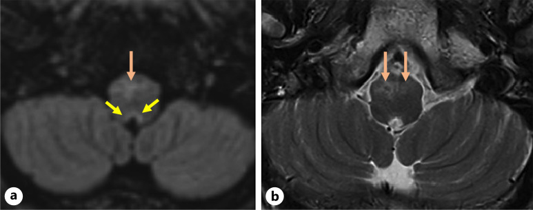
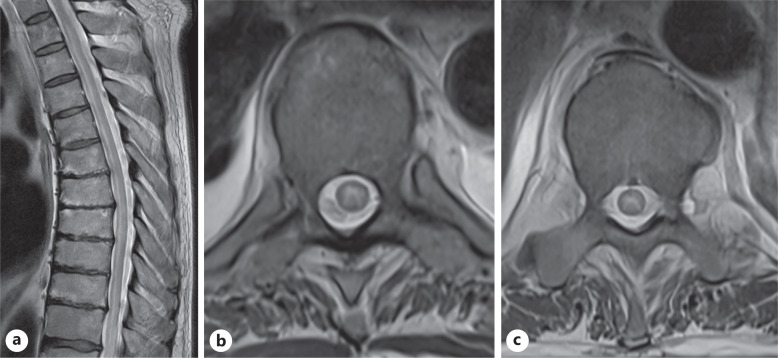
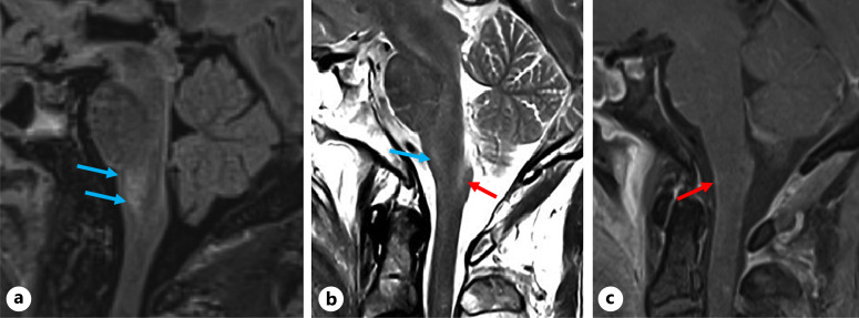
Case presentation: A previously healthy man in his 70s presented with headache, fever, and confusion, followed by a seizure and persistent hiccoughs. MRI revealed brainstem involvement and extensive transverse myelitis. Cerebrospinal fluid (CSF) analysis showed inflammatory features, but testing for AQP4, MOG, and GFAP antibodies was initially negative. He was treated with intravenous corticosteroids and plasma exchange, after which serum GFAP-IgG was weakly positive, though CSF remained negative. His condition improved with immunotherapy, but significant lower limb weakness persisted. Based on clinical and radiological findings, we hypothesize that tanycytes - specialized glial cells in the area postrema - may be an additional immune target in GFAP encephalitis.
Conclusion: This case highlights a seronegative encephalomyelitis syndrome with area postrema involvement, possibly implicating glial cells beyond astrocytes. Further studies are needed to explore the role of tanycytes in autoimmune neuroinflammation.
期刊介绍:
This new peer-reviewed online-only journal publishes original case reports covering the entire spectrum of neurology. Clinicians and researchers are given a tool to disseminate their personal experience to a wider public as well as to review interesting cases encountered by colleagues all over the world. To complement the contributions supplementary material is welcomed. The reports are searchable according to the key words supplied by the authors; it will thus be possible to search across the entire growing collection of case reports with universally used terms, further facilitating the retrieval of specific information. Following the open access principle, the entire contents can be retrieved at no charge, guaranteeing easy access to this valuable source of anecdotal information at all times.

 求助内容:
求助内容: 应助结果提醒方式:


